Anatomical Dental Chart
Anatomical Dental Chart - For primary teeth, most dentists in united states use a modified version of the universal numbering system, with each primary tooth assigned a letter (from a to t) instead of a. Primary (baby or deciduous) teeth names & numbers. In this page, we are going to study each one of the above types, learn how they are numbered, and understand the various anatomical parts of teeth. There are separate teeth number charts for adults as well as babies. Web a free dental library of interactive 3d models for dental education including dental anatomy, occlusion, prosthodontics and endodontics. The canines have the largest and strongest roots. Web detail the differences in tooth diagrams, including anatomical and geometric charting using the universal/national numbering system. The development, appearance, and classification of teeth fall within its purview. Web dental anatomy is a field of anatomy dedicated to the study of human tooth structures. This is the part of your tooth that you can see — the portion above your gums. Web atlas of dental anatomy: Web browse our selection of dental charts and posters, ideal for learning or teaching anatomy and physiology of the mouth and teeth, including dental pathologies and how to maintain good oral health. Web dental anatomy is a field of anatomy dedicated to the study of human tooth structures. The anterior teeth consist of the incisors. Web an overview of dental anatomy. Each tooth has a crown and a root. For primary teeth, most dentists in united states use a modified version of the universal numbering system, with each primary tooth assigned a letter (from a to t) instead of a. Web anatomy of the teeth laminated anatomical chart. Web a free dental library of interactive. The numbering system shown is the one most commonly used in the united states. Web atlas of dental anatomy: You can’t see the root because your gums cover it. Web the teeth are divided into four quadrants within the mouth, with the division occurring between the upper and lower jaws horizontally and down the midline of the face vertically. Web. Web detail the differences in tooth diagrams, including anatomical and geometric charting using the universal/national numbering system. Web imaging planes and reconstructions were chosen according to those used the most often in daily routine: Web a free dental library of interactive 3d models for dental education including dental anatomy, occlusion, prosthodontics and endodontics. In this page, we are going to. In this page, we are going to study each one of the above types, learn how they are numbered, and understand the various anatomical parts of teeth. The development, appearance, and classification of teeth fall within its purview. When identifying teeth and referring to specific areas of a tooth, it is necessary to utilize named surfaces and directions designated according. Web anatomy of the teeth laminated anatomical chart. An inner pulp contains blood vessels, lymphatics, and nerves, surrounded by the hard but porous dentin, which is sensitive to touch and to temperature changes. Web imaging planes and reconstructions were chosen according to those used the most often in daily routine: Periodontal charting, which is a part of your dental. This. Web dental anatomy is a field of anatomy dedicated to the study of human tooth structures. Each type of tooth has a specific function, including. A tooth consists of two main structures: Web dental charting is a process in which your dental healthcare professional lists and describes the health of your teeth and gums. Web buy dental anatomy posters and. Web an overview of dental anatomy. This is the part of your tooth that you can see — the portion above your gums. You can’t see the root because your gums cover it. The permanent dentition is composed of 32 teeth with 16 in each arch. Web what’s the anatomy of a tooth? Web the anterior teeth are the twelve teeth in the front of the mouth, while the posterior teeth are the teeth in the back of the mouth. A tooth consists of two main structures: Each tooth has a crown and a root. You can’t see the root because your gums cover it. This diagram helps us learn the names of. The labels right and left on the charts in this article correspond to the patient's right and left, respectively. Each tooth has a crown and a root. Educate students and patients with these anatomical wall charts. Web the teeth are divided into four quadrants within the mouth, with the division occurring between the upper and lower jaws horizontally and down. The canines have the largest and strongest roots. For primary teeth, most dentists in united states use a modified version of the universal numbering system, with each primary tooth assigned a letter (from a to t) instead of a. Web there are five teeth in each quadrant, composed of two incisors (central and lateral), a canine, and two molars. Choose the appropriate surfaces and classifications for existing and planned restorations. The patient's right side appears on the left side of the chart, and the patient's left side appears on the right side of the chart. You can’t see the root because your gums cover it. The permanent dentition is composed of 32 teeth with 16 in each arch. The numbering system shown is the one most commonly used in the united states. Web this article will explain the different types of human teeth, their function, and how they're charted by dental professionals to help track of changes in your dental health. (the function of teeth as they contact one another falls elsewhere, under dental occlusion.) Learn about the types of teeth in a fast and efficient way using our interactive tooth identification quizzes and labeled diagrams. There are dental charts showing disorders of the jaw and other diseases of the dental structure. This is the part of your tooth that you can see — the portion above your gums. Web left and right on the teeth chart correspond to the patient's left and right respectively (patient's view). The development, appearance, and classification of teeth fall within its purview. Web dental anatomy is defined here as, but is not limited to, the study of the development, morphology, function, and identity of each of the teeth in the human dentitions, as well as the way in which the teeth relate in shape, form, structure, color, and function to the other teeth in the same dental arch and to the teeth in the opposing arch.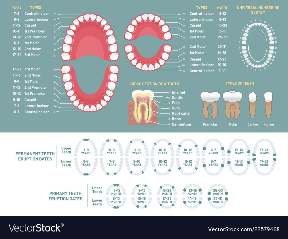
Tooth anatomy chart orthodontist human teeth loss Vector Image

The Different Types of Teeth Gentle Dentist
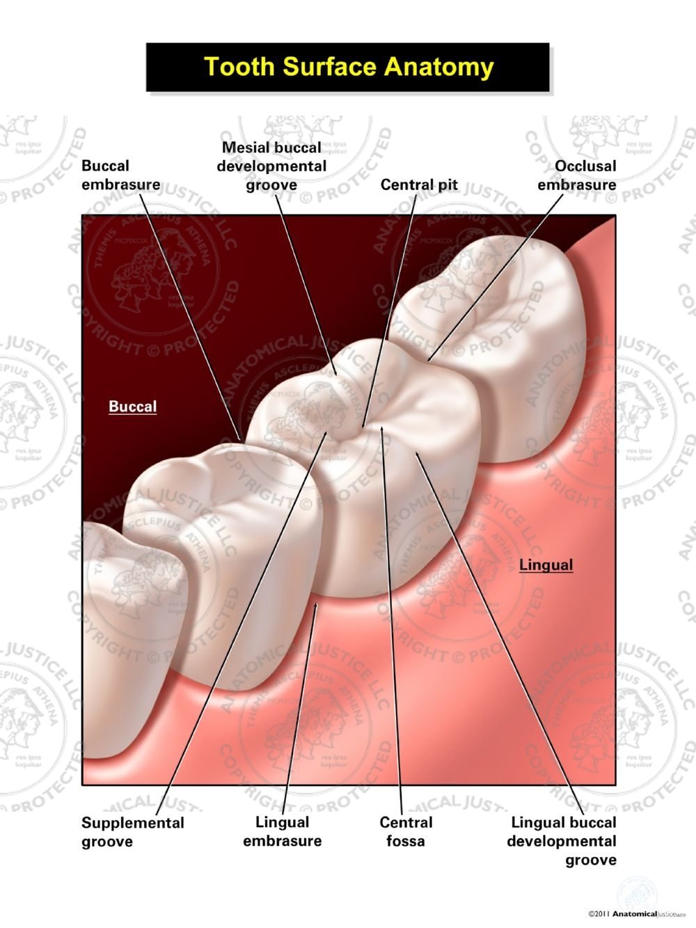
Tooth Surface Anatomy

Tooth Number Chart to Identify Primary Teeth Eruption Charts
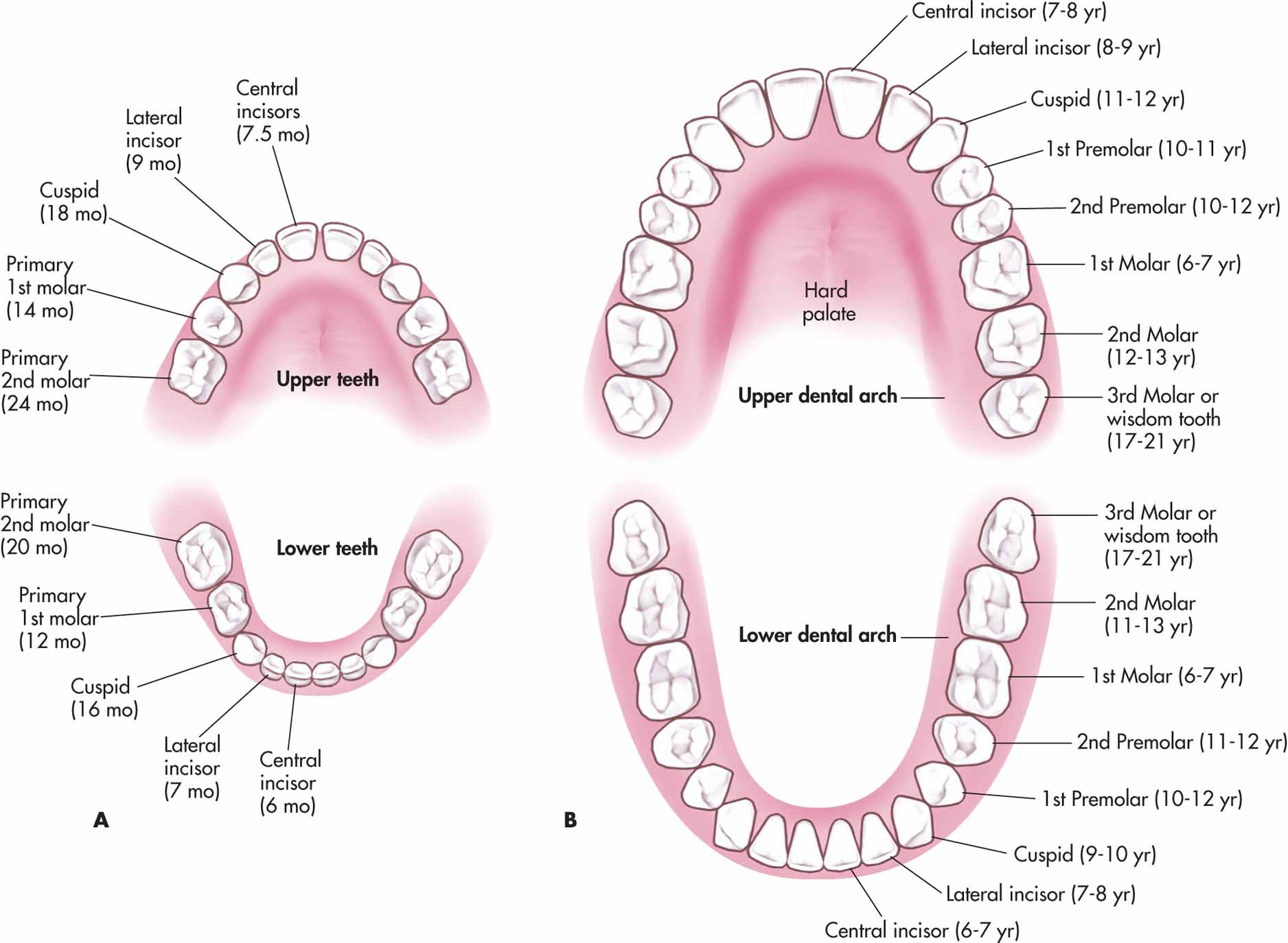
Deciduous And Permanent Teeth and Structure of a Tooth Earth's Lab

Printable Tooth Chart With Numbers And Letters Web A Tooth Numbering
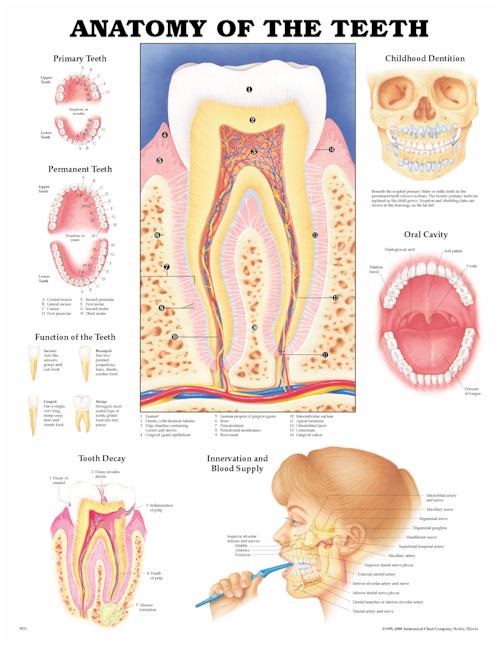
Tooth Chart (Anatomical Charts) Dental Product Pearson Dental
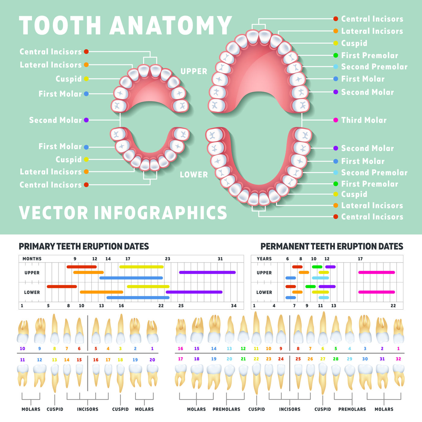
tooth number diagrams

Child and Adult Dentition (Teeth) Structure Primary Permanent
Dental anatomy reference_guide
The Primary Teeth Begin To Erupt At 6 Months Of Age.
Web A Free Dental Library Of Interactive 3D Models For Dental Education Including Dental Anatomy, Occlusion, Prosthodontics And Endodontics.
Prefer To Learn By Doing?
Teeth Also Have Number/Letter Designations.
Related Post: