Anatomical Tooth Chart
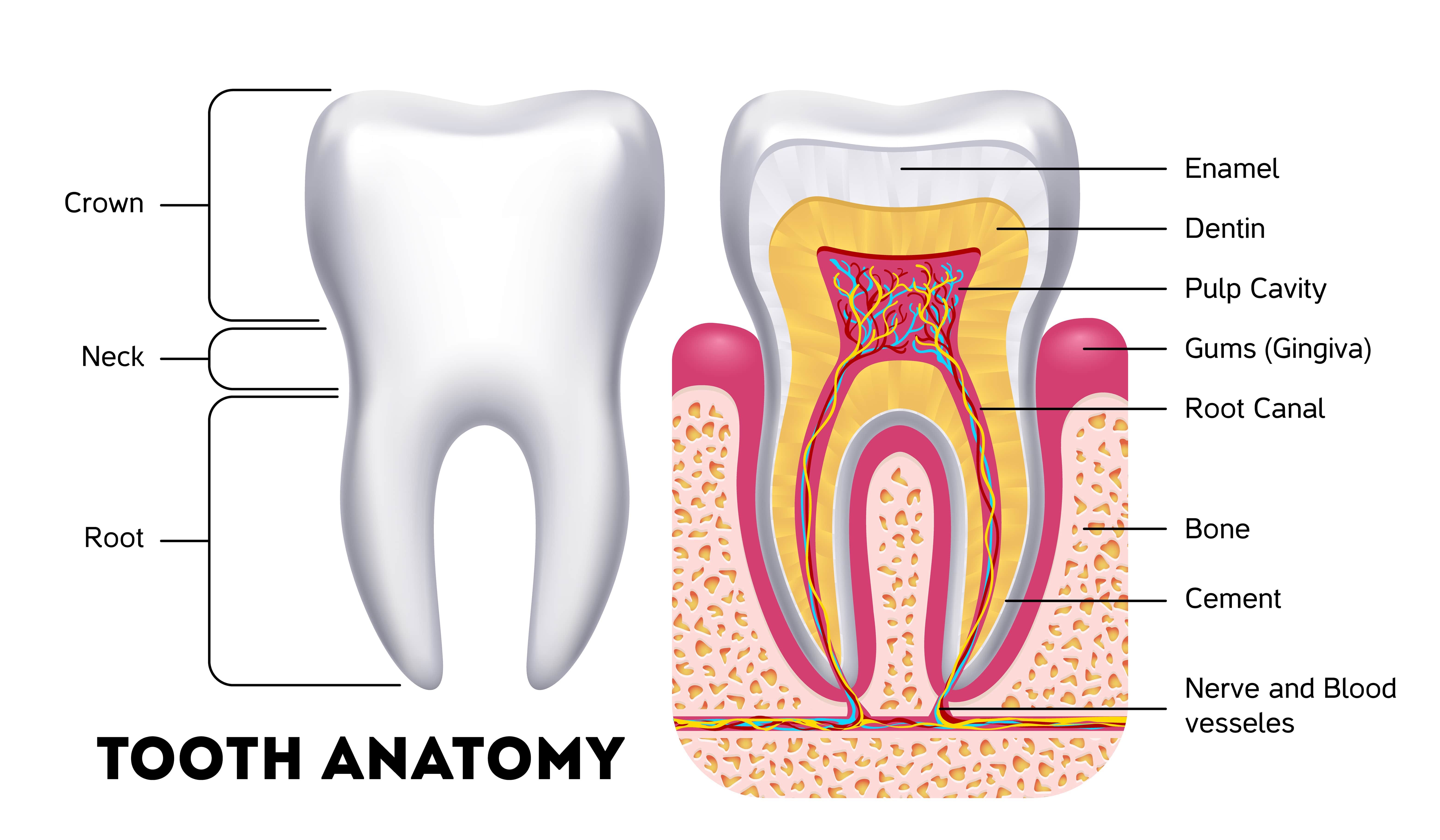
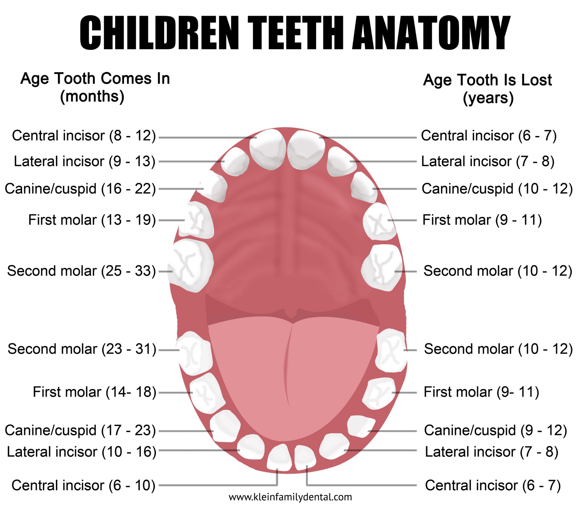
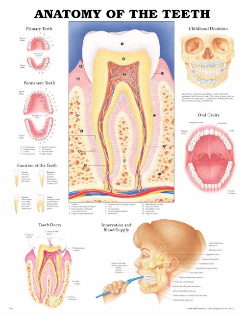
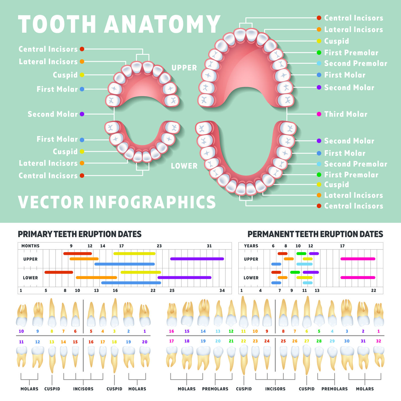
Anatomical Tooth Chart - Brightly colored, user friendly chart covering the anatomy of the teeth. The crown of the tooth is what is visible in the oral cavity, and the root of the tooth is embedded into the bony ridge of the upper and lower jaws called the alveolar process via attachment to the periodontal ligament. Web a free dental library of interactive 3d models for dental education including dental anatomy, occlusion, prosthodontics and endodontics. Your teeth play a big role in digestion. Web left and right on the teeth chart correspond to the patient's left and right respectively (patient's view). Most adults have 32 permanent teeth, including eight incisors, four canines, eight premolars and 12 molars. There are four main types of teeth in humans, shown labelled here. The outer surface is a thin layer of enamel which covers the inner dentin. There are separate teeth number charts for adults as well as babies. The crown and the root. Web anatomy of the teeth anatomical chart. Temporomandibular joint posters and much more are also available. Web the anatomy of a tooth divides into two main sections: Like all teeth, canines have a crown (the part above the gum), a neck and a root (the part inside the bone). Web left and right on the teeth chart correspond to the. For example, in posterior teeth, mandibular molar, the five surfaces are buccal, occlusal, lingual, mesial, and distal surfaces. Web the anatomy of a tooth divides into two main sections: Web dental charts are normally arranged from the viewpoint of a dental practitioner facing a patient. Primary (baby or deciduous) teeth names & numbers. Web left and right on the teeth. Web the teeth are categorized as incisors, canines, premolars, and molars and conventionally are numbered beginning with the maxillary right third molar (see figure identifying the teeth). The outer surface is a thin layer of enamel which covers the inner dentin. Fully labeled illustrations of the teeth with dental terminology (orientation, surfaces, cusps, roots numbering systems) and detailed images of. There are dental charts showing disorders of the jaw and other diseases of the dental structure. Teeth also have number/letter designations. Fully labeled illustrations of the teeth with dental terminology (orientation, surfaces, cusps, roots numbering systems) and detailed images of each permanent tooth. Most adults have 32 permanent teeth, including eight incisors, four canines, eight premolars and 12 molars. Prefer. The large central image shows a detailed cross. Web atlas of dental anatomy: Web the teeth are categorized as incisors, canines, premolars, and molars and conventionally are numbered beginning with the maxillary right third molar (see figure identifying the teeth). Web left and right on the teeth chart correspond to the patient's left and right respectively (patient's view). Web we’ll. Each type of tooth has a specific function, including biting, chewing, and grinding up food. Your teeth play a big role in digestion. The crown of the tooth is what is visible in the oral cavity, and the root of the tooth is embedded into the bony ridge of the upper and lower jaws called the alveolar process via attachment. Dental anatomy is a field of anatomy dedicated to the study of tooth structure. We’ll also go over some common conditions that can affect your teeth, and we’ll list common symptoms to watch for. Following is a brief description of this topic. Primary (baby or deciduous) teeth names & numbers. Your teeth play a big role in digestion. Web atlas of dental anatomy: The anterior teeth are the twelve teeth in the front of the mouth, while the posterior teeth are the teeth in the back of the mouth. Web a teeth chart is a simple drawing or illustration of your teeth with names, numbers, and types of teeth. Teeth are made up of. The primary teeth begin. Web image from visible body suite. Dental anatomy is a field of anatomy dedicated to the study of tooth structure. The large central image shows a detailed cross. We’ll also go over some common conditions that can affect your teeth, and we’ll list common symptoms to watch for. Web dental anatomy is a field of anatomy dedicated to the study. Dental anatomy is a field of anatomy dedicated to the study of tooth structure. The large central image shows a detailed cross. Web the 4 main tooth types are incisors, canines, premolars, and molars. The names are given to these surfaces according to their position and use. Web dental anatomy is a field of anatomy dedicated to the study of. Web we’ll go over the anatomy of a tooth and the function of each part. Web surfaces of the teeth. There are dental charts showing disorders of the jaw and other diseases of the dental structure. This leaves up to eight adult teeth in each quadrant and separates the opposing pairs within the same alveolar bone as well as their counterparts in the opposing jaw. Look no further than our dental anatomy quizzes and tooth diagrams. Each type of tooth has a specific function, including biting, chewing, and grinding up food. Teeth also have number/letter designations. We’ll also go over some common conditions that can affect your teeth, and we’ll list common symptoms to watch for. There are separate teeth number charts for adults as well as babies. There are four main types of teeth in humans, shown labelled here. These teeth are referred to as letters a, b, c, d and e. Web humans have four canine teeth, two maxillary canine teeth (left and right) and two mandibular canine teeth (left and right). The anterior teeth are the twelve teeth in the front of the mouth, while the posterior teeth are the teeth in the back of the mouth. There are five teeth in each quadrant, composed of two incisors (central and lateral), a canine, and two molars. For example, in posterior teeth, mandibular molar, the five surfaces are buccal, occlusal, lingual, mesial, and distal surfaces. For primary teeth, most dentists in united states use a modified version of the universal numbering system, with each primary tooth assigned a letter (from a to t) instead of a.
Dental anatomy reference_guide
Anatomy Of The Teeth Anatomical Chart Poster Prints Images and Photos

Printable Tooth Chart With Numbers And Letters Web A Tooth Numbering
Pediatric Tooth Chart — Klein Family Dental

Child and Adult Dentition (Teeth) Structure Primary Permanent
Tooth Chart (Anatomical Charts) Dental Product Pearson Dental

Tooth Number Chart to Identify Primary Teeth Eruption Charts
tooth number diagrams
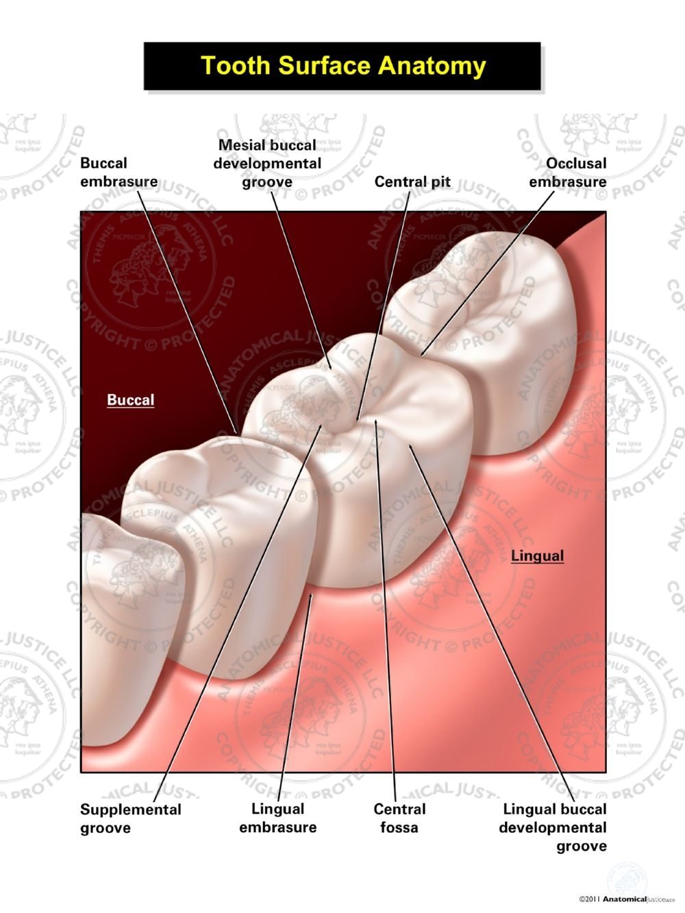
Tooth Surface Anatomy

The Different Types of Teeth Gentle Dentist
The Large Central Image Shows A Detailed Cross.
Web The 4 Main Tooth Types Are Incisors, Canines, Premolars, And Molars.
Web Image From Visible Body Suite.
The Two Types Are The Central Incisors And Lateral Incisors.
Related Post: