Canine Anatomy Chart
Canine Anatomy Chart - A brief outline of diagnostic, In addition to the world’s most segmented dog anatomy, the table vet also includes a diverse library of animal cases. Surely you're familiar with common features such as the legs, eyes, and tail… but how about the loin or the hock? The size of the tail, and therefore the number of bones in a dog, is dependent on the length of their tail. Web 740 anatomical terms have been labeled, organized in different sections : Web here are presented scientific illustrations of the canine skeleton, with the main dog's bones and its structures displayed from different anatomical standard views (cranial, caudal, lateral, medial, dorsal, palmar.). In this article, you will learn the location of different organs from the different systems (like skeletal, digestive, respiratory, urinary, cardiovascular, endocrine, nervous, and special sense) of a dog with their important anatomical features. Web featuring the world’s most comprehensive canine cadaver, the anatomage table vet promises to bring animal bodies back to life and visualize them in 3d accuracy. Cannes film festival (un certain regard) cast: Each illustration in the atlas has been drawn by professional medical illustrators. Their front legs are attached to their shoulders and are used for steering and braking, while their hind legs are attached to their hips and are used for propulsion. Seven cervical vertebrae, thirteen thoracic vertebrae, seven lumbar vertebrae and three sacral vertebrae. Web their skeleton includes their skull, spine, ribcage and limbs. Posterior view of the skeleton with lateral view. Some of the different canine joints are labeled. Web understand dog anatomy with our canine charts and models, including skeletons and pathology models. Details of structures vary tremendously from breed to breed, more than in any other animal species, wild or domesticated, as dogs are highly variable in height and weight. Seven cervical vertebrae, thirteen thoracic vertebrae, seven lumbar vertebrae. Web dog anatomy comprises the anatomical study of the visible parts of the body of a domestic dog. Web the following canine anatomy illustrations offer a look at various systems within the dog's body. Web directional terms and anatomic planes. Muscle, organ and skeletal anatomy). Web this ct of a spayed female dog’s whole body was performed after injection of. Details of structures vary tremendously from breed to breed, more than in any other animal species, wild or domesticated, as dogs are highly variable in height and weight. Surely you're familiar with common features such as the legs, eyes, and tail… but how about the loin or the hock? Although these pictures are fairly basic, they still provide insight that. A dog’s skeleton is divided into the axial skeleton (skull, vertebrae, ribs, and sternum) and the appendicular skeleton (limbs, shoulder girdle, and pelvic girdle). Some of the different canine joints are labeled. Web their skeleton includes their skull, spine, ribcage and limbs. Web understand dog anatomy with our canine charts and models, including skeletons and pathology models. Web here are. Their front legs are attached to their shoulders and are used for steering and braking, while their hind legs are attached to their hips and are used for propulsion. In this article, you will learn the location of different organs from the different systems (like skeletal, digestive, respiratory, urinary, cardiovascular, endocrine, nervous, and special sense) of a dog with their. Can you name all of the parts of a dog? Let’s review the anatomical terms used to describe the parts of the dogs, starting from head to tail. Web dog anatomy details the various structures of canines (e.g. Web anatomical terms you should know. Web anatomy atlas of the canine general anatomy: Positional and directional terms, general terminology and anatomical orientation are also illustrated. Dogs have four legs that are designed to help them move quickly and efficiently. Web gain a comprehensive understanding of your dog's health with our veterinary guide to cat anatomy complete with diagrams, images and simple explanations. A brief outline of diagnostic, A dog’s skeleton is divided into. Cannes film festival (un certain regard) cast: Web atlas of the canine anatomy based on veterinary anatomy diagrams and medical images (radiographs, ct, mri, endoscopy). Web here are presented scientific illustrations of the canine skeleton, containing the main joints of the dog and its structures from different standard anatomical views (cranial, caudal, lateral, medial, dorsal, palmar.). Web the anatomy of. Surely you're familiar with common features such as the legs, eyes, and tail… but how about the loin or the hock? The detailing of these structures changes based on dog breed due to the huge variation of size in dog breeds. The images are transverse sections of the whole body and a. General anatomy of the skull. Web their skeleton. Web anatomical terms you should know. Web 740 anatomical terms have been labeled, organized in different sections : Web this ct of a spayed female dog’s whole body was performed after injection of a contrast medium by delphine rault, dvm, dipl. Muscle, organ and skeletal anatomy). Details of structures vary tremendously from breed to breed, more than in any other animal species, wild or domesticated, as dogs are highly variable in height and weight. Fully labeled illustrations and diagrams of the dog (skeleton, bones, muscles, joints, viscera, respiratory system, cardiovascular system). The following paragraphs explain all these aspects in brief, along with diagrams, which will help you understand them better. A brief outline of diagnostic, Web the following canine anatomy illustrations offer a look at various systems within the dog's body. Web this veterinary anatomy module contains 608 illustrations on the canine myology. Web featuring the world’s most comprehensive canine cadaver, the anatomage table vet promises to bring animal bodies back to life and visualize them in 3d accuracy. Web this is why animalwised brings you this dog anatomy guide where we look at the general categories for muscles, bones and organs of dogs. Their front legs are attached to their shoulders and are used for steering and braking, while their hind legs are attached to their hips and are used for propulsion. Web labeled view of the internal organs. Dogs have approximately 320 bones in their bodies, depending on the breed. Web atlas of the canine anatomy based on veterinary anatomy diagrams and medical images (radiographs, ct, mri, endoscopy).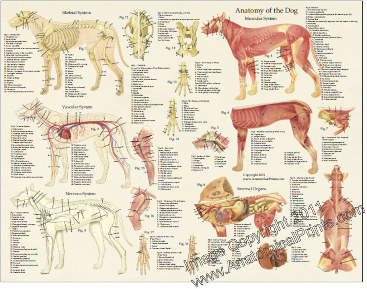
Dog Anatomy Laminated Poster Clinical Charts and Supplies
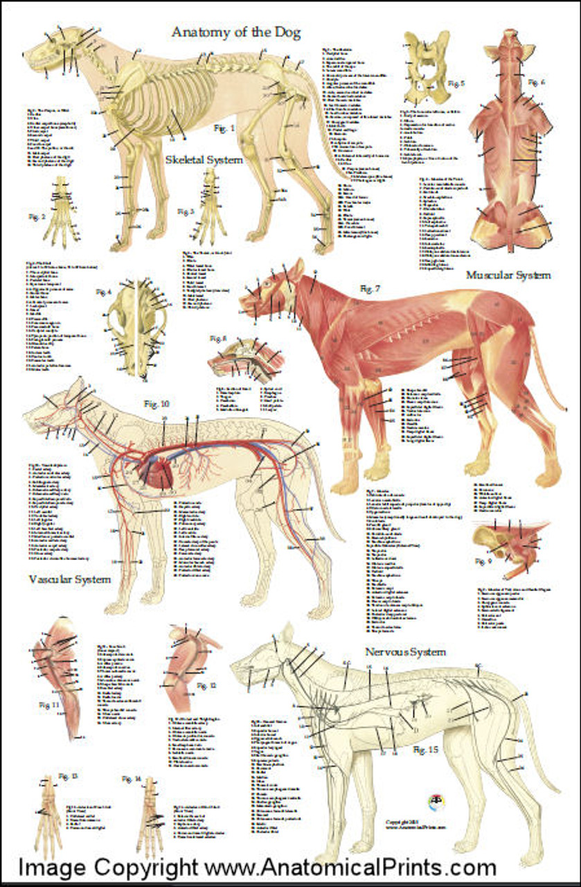
Dog Anatomy Poster 24 x 36 Clinical Charts and Supplies

Canine Internal Anatomy Chart Poster Laminated studiosixsound.co.za
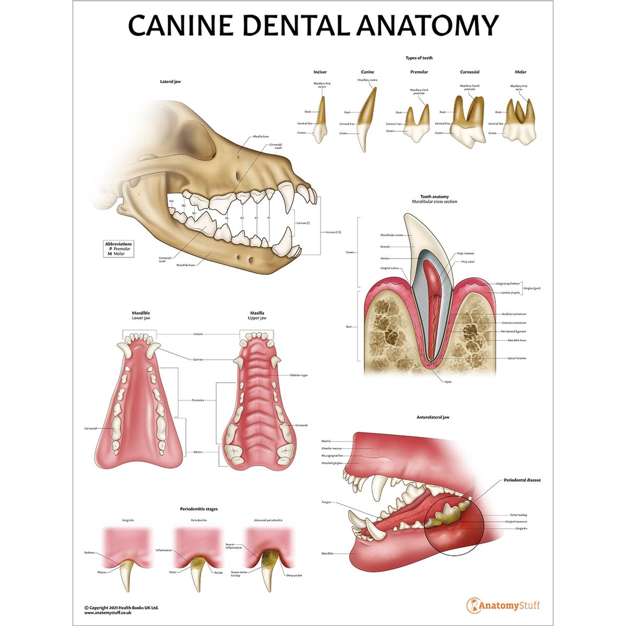
Canine Anatomy Models, Charts & Simulators Dog Skeleton Page 3

the anatomy of a dog's body and its major skeletal systems are shown in
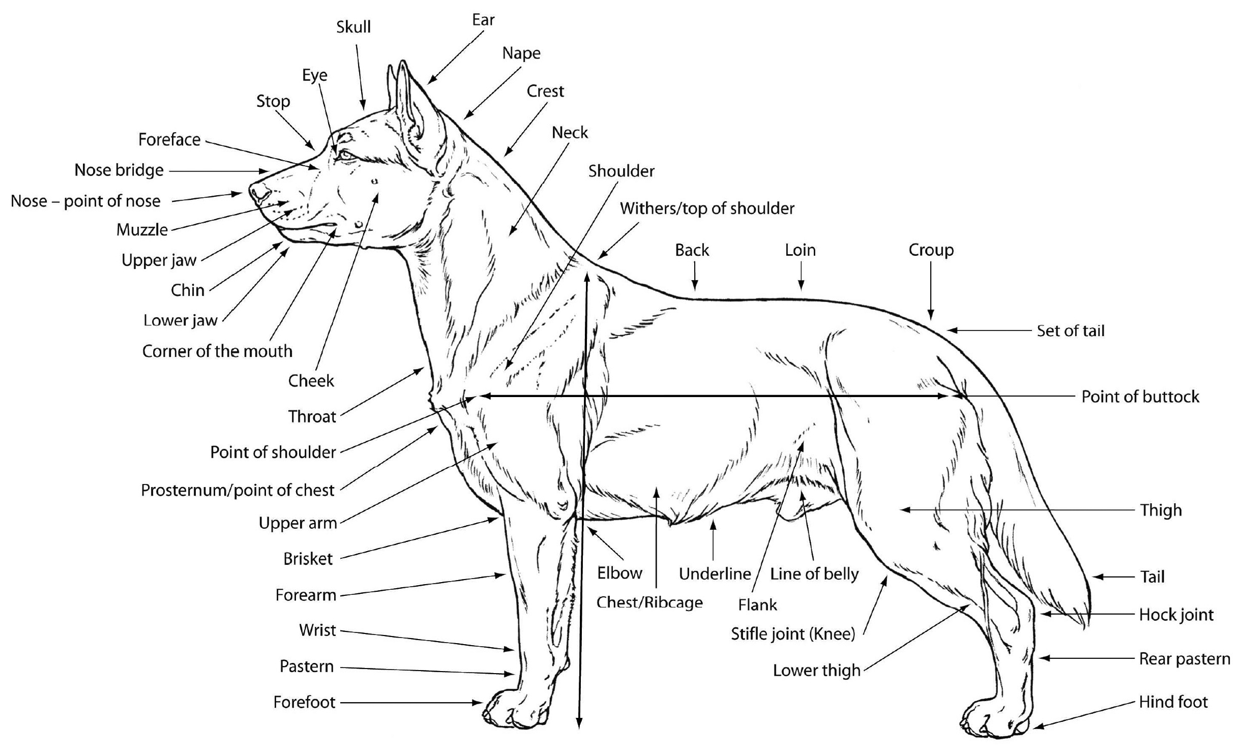
M. Douglas Wray Dog Anatomy
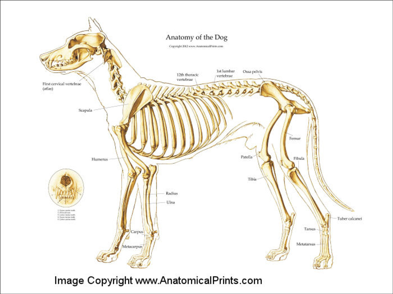
Canine Skeleton Poster Clinical Charts and Supplies

Canine Anatomy, Complete Set of 3 Charts. Buy The Set and Save! Amazon
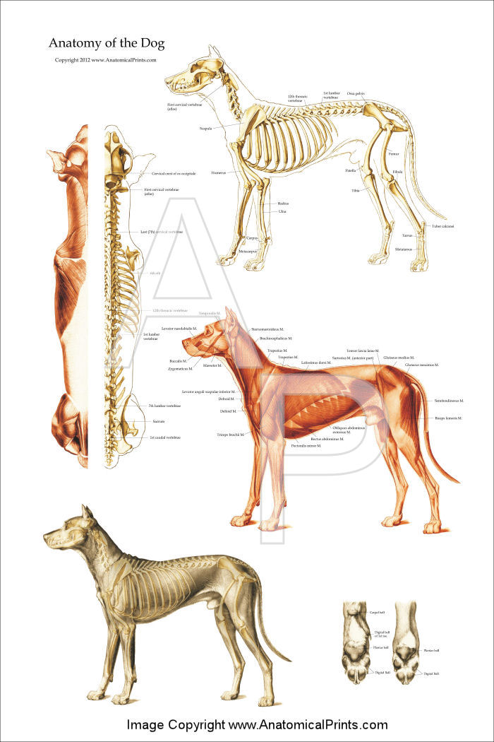
Dog Anatomical Chart Bones and Muscles
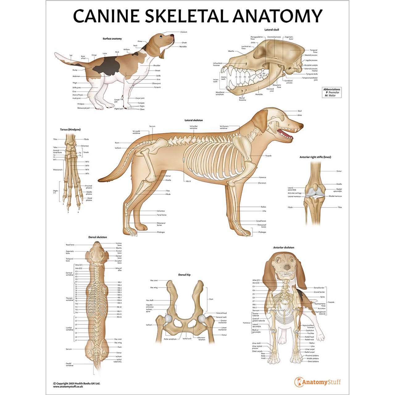
Canine Skeletal Anatomy Laminated Chart Dog Skeleton Poster
Seven Cervical Vertebrae, Thirteen Thoracic Vertebrae, Seven Lumbar Vertebrae And Three Sacral Vertebrae.
Web Gain A Comprehensive Understanding Of Your Dog's Health With Our Veterinary Guide To Cat Anatomy Complete With Diagrams, Images And Simple Explanations.
In This Article, You Will Learn The Location Of Different Organs From The Different Systems (Like Skeletal, Digestive, Respiratory, Urinary, Cardiovascular, Endocrine, Nervous, And Special Sense) Of A Dog With Their Important Anatomical Features.
Can You Name All Of The Parts Of A Dog?
Related Post: