Chart Of Nerves In Back
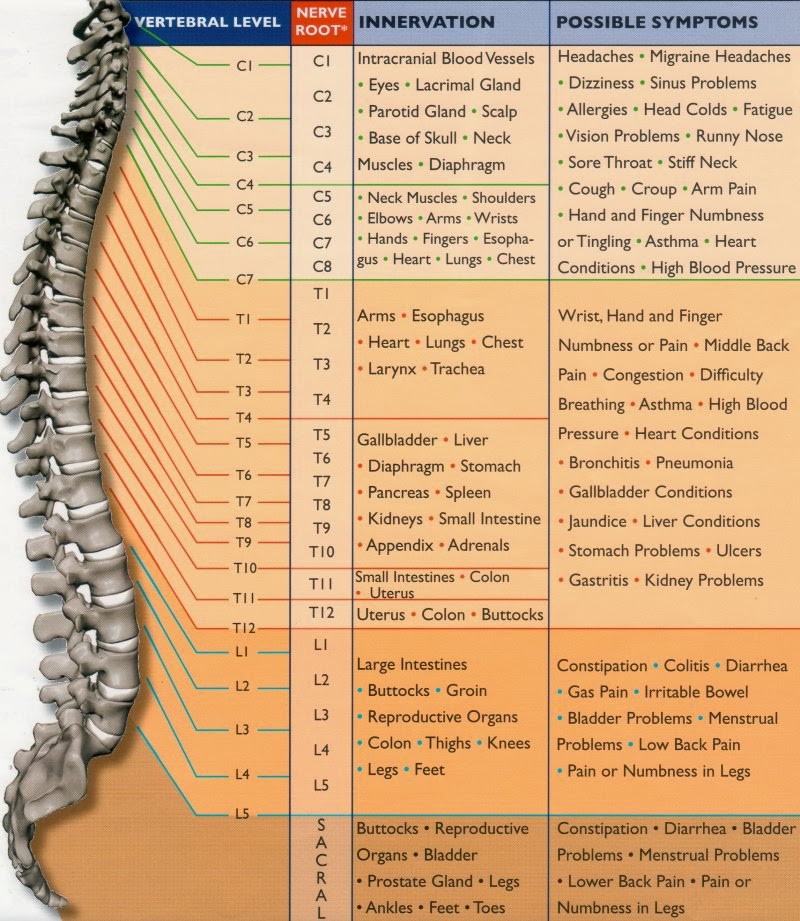
Chart Of Nerves In Back - Web there are 31 pairs of spinal nerves: The cauda equina is formed from the spinal nerves which arise from the end of the spinal cord. There are small sensory nerves along these joints whose only job is to tell the brain how the joint feels. There are things you can do to help ease the pain. Web learn the anatomy of the spinal nerves, including their roots, components and functions faster and more efficiently with this comprehensive article. Spinal pain can arise from problems in the bones, nerves, or other soft tissues. As these nerves descend toward the thighs, they form two networks of crossed nerves known as the lumbar plexus and sacral plexus. Your spine has 33 stacked vertebrae (small bones) that form the spinal canal. Typical anatomical problems that cause back pain. Learn about their role in transmitting signals and their impact on lower limb mobility. Web the relevant anatomy of the innervation of the musculature of the back by the spinal nerves is centered around the lumbar spinal nerves, peripheral nerves of the lumbar plexus, spinal cord, and lumbar vertebral column. Web explore the anatomy and functions of lumbar spinal nerves. Web health a to z. Back pain can have many causes. There are small. At the level of the l2 vertebra, the spinal cord becomes known as the cauda equina. But by driving social mobility and boosting security, the pm’s plan will help us chart our way in an uncertain. Web the spinal cord has five sections of spinal nerves branching off. Web many nerves come from the spinal cord, pass through foramina (holes). The spinal cord and peripheral nerves. On the chart below you will see 4 columns (vertebral level, nerve root, innervation, and possible symptoms). Web the nerves of the leg and foot arise from spinal nerves connected to the spinal cord in the lower back and pelvis. At the level of the l2 vertebra, the spinal cord becomes known as the. Web the spine’s four sections, from top to bottom, are the cervical (neck), thoracic (abdomen,) lumbar (lower back), and sacral (toward tailbone). Web these nerves are located at the cervical (neck), thoracic (upper back), lumbar (lower back), sacral (sacrum, which forms part of the pelvis), and coccygeal (tailbone) levels. Many of the intricate structures in the spine can lead to. At the level of the l2 vertebra, the spinal cord becomes known as the cauda equina. Web there are seven cervical vertebrae at the top, followed by 11 thoracic vertebrae, five lumbar vertebrae at the lower back, and five fused vertebrae at the bottom to create the sacrum. With student loans, that extra on the top isn't so little right. The lumbar plexus forms in the lower back from the merger of spinal nerves l1 through l4. Web explore the anatomy and functions of lumbar spinal nerves. Web health a to z. But by driving social mobility and boosting security, the pm’s plan will help us chart our way in an uncertain. Compared with other spine vertebrae, your lumbar vertebrae. Spinal pain can arise from problems in the bones, nerves, or other soft tissues. Throughout the spine, intervertebral discs made of. Web the nerves of the leg and foot arise from spinal nerves connected to the spinal cord in the lower back and pelvis. Web explore the anatomy and functions of lumbar spinal nerves. The spinal cord and peripheral nerves. The symptoms of a pinched. On the chart below you will see 4 columns (vertebral level, nerve root, innervation, and possible symptoms). Compared with other spine vertebrae, your lumbar vertebrae are. Each pair of spinal nerves are dedicated to certain regions of the body. At the level of the l2 vertebra, the spinal cord becomes known as the cauda equina. Web there are 31 pairs of spinal nerves: Web many nerves come from the spinal cord, pass through foramina (holes) formed by notches of 24 vertebrae in the movable spinal column, and innervate or supply specific areas and parts of the body.2 whenever specific areas or parts of the body are malfunctioning, generalized and/or specific symptoms are possible. Web there. Back pain can have many causes. Web the nerves of the leg and foot arise from spinal nerves connected to the spinal cord in the lower back and pelvis. At the level of the l2 vertebra, the spinal cord becomes known as the cauda equina. Web explore the anatomy and functions of lumbar spinal nerves. The spinal cord serves as. The symptoms of a pinched. There are small sensory nerves along these joints whose only job is to tell the brain how the joint feels. The spinal canal is a tunnel that houses your spinal cord and nerves, protecting them from injury. 8 cervical, 12 thoracic, 5 lumbar, 5 sacral, and 1 coccygeal, named according to their corresponding vertebral levels. Web the spinal cord has five sections of spinal nerves branching off. These nerves are the primary source of pain signals coming from the joints of the back. Web there are 31 pairs of spinal nerves, forming nerve roots that branch from your spinal cord. Web these nerves are located at the cervical (neck), thoracic (upper back), lumbar (lower back), sacral (sacrum, which forms part of the pelvis), and coccygeal (tailbone) levels. The lumbar plexus forms in the lower back from the merger of spinal nerves l1 through l4. On the chart below you will see 4 columns (vertebral level, nerve root, innervation, and possible symptoms). Web the spine’s four sections, from top to bottom, are the cervical (neck), thoracic (abdomen,) lumbar (lower back), and sacral (toward tailbone). At the level of the l2 vertebra, the spinal cord becomes known as the cauda equina. As these nerves descend toward the thighs, they form two networks of crossed nerves known as the lumbar plexus and sacral plexus. The spinal cord serves as the central pathway for transmitting sensory and motor signals between the brain and the body through specific. The peripheral nerves are responsible for sensations and muscle movements. Web many nerves come from the spinal cord, pass through foramina (holes) formed by notches of 24 vertebrae in the movable spinal column, and innervate or supply specific areas and parts of the body.2 whenever specific areas or parts of the body are malfunctioning, generalized and/or specific symptoms are possible.
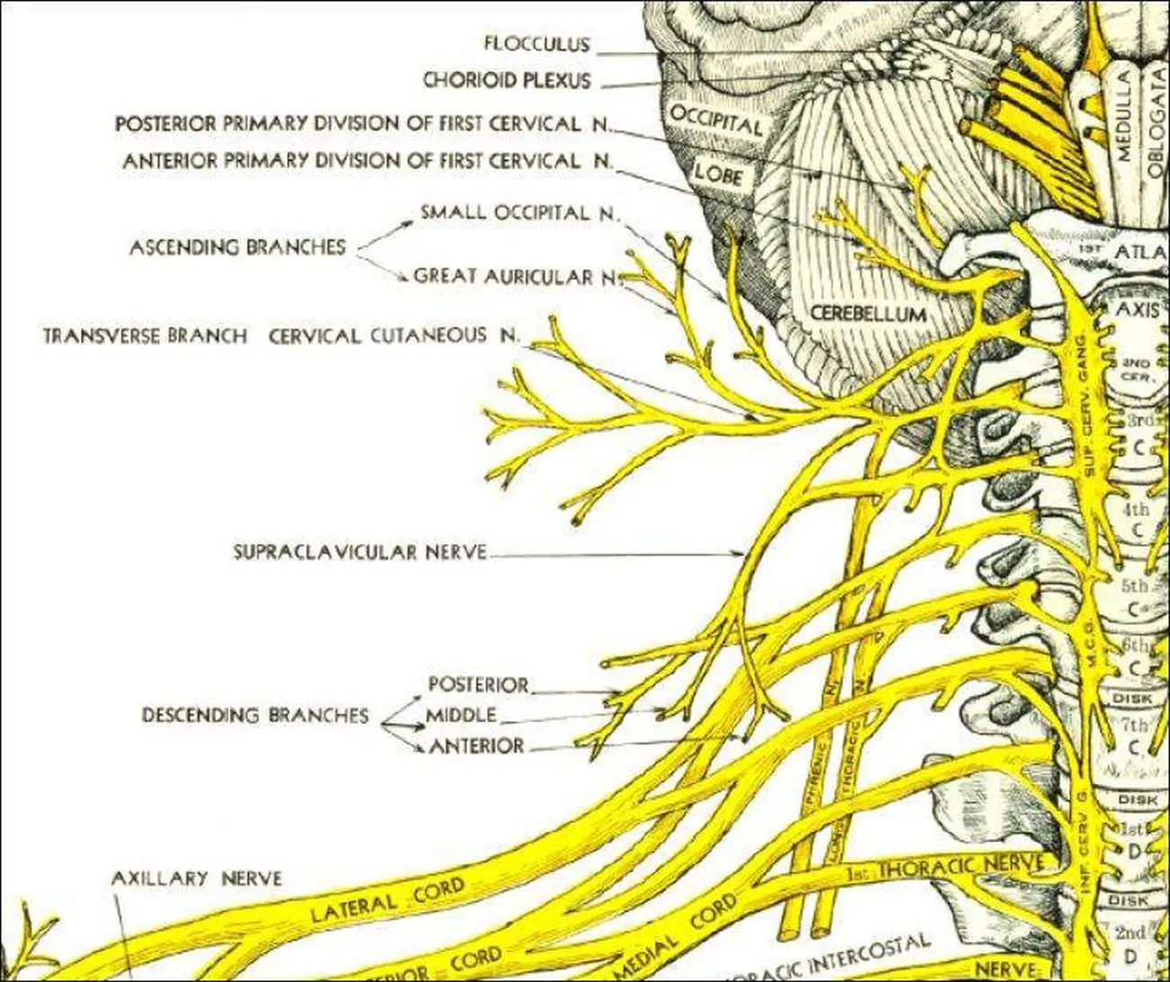
the spiral nerve function is shown in this manual for students to learn

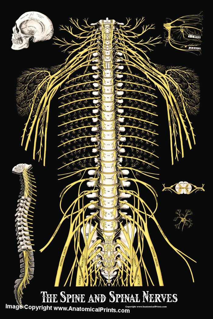
The Spine and Spinal Nerves Poster Clinical Charts and Supplies

Anatomy Of Organs In The Body Back View Michael Heathcaldwell M.arch

Spinal Nerve Chart medschool doctor medicalstudent Image Credits

The Spinal Nerves Chart

Spinal Nerves Anatomical Chart Spine and Cranial Nervous System

Spinal nerve function. OT Physical therapy, Medicine, Neurology

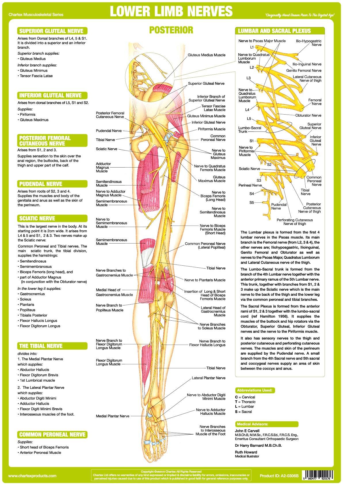
Lower Back Nerves Body Diagram Sciatica pain vector illustration

Nervous System Anatomy Posters Set of 6

Chart Of Nerves In Back
Web The Relevant Anatomy Of The Innervation Of The Musculature Of The Back By The Spinal Nerves Is Centered Around The Lumbar Spinal Nerves, Peripheral Nerves Of The Lumbar Plexus, Spinal Cord, And Lumbar Vertebral Column.
Web Explore The Anatomy And Functions Of Lumbar Spinal Nerves.
Learn About Their Role In Transmitting Signals And Their Impact On Lower Limb Mobility.
In General, The Spinal Cord Consists Of Gray And White Matter.
Related Post: