Deltoid Muscle Diagram Drawing
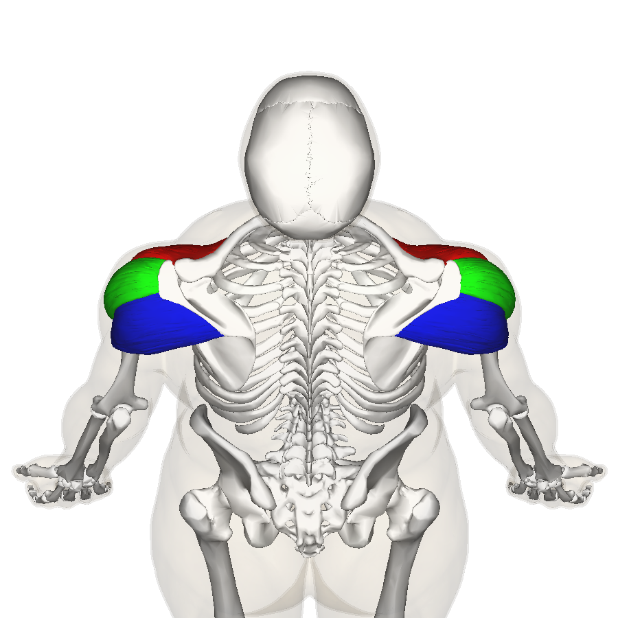
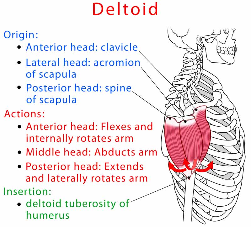
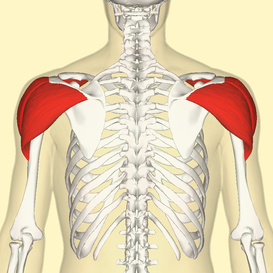
Deltoid Muscle Diagram Drawing - Web in this captivating diagram video, we delve into the depths of one of the most prominent and powerful muscles in the human body. Another common mistake is drawing it. Web the deltoid muscle is known for its origin’s distinctive ‘u’ shape, spanning across the clavicle, acromion, and the spine of the scapula. Web let’s now draw in the shoulders on top of the foundation in these diagrams. Web front and back view isolated vector illustrations on white background. [1] [2] the shoulder girdle is composed of the following osseous components. The functions of the deltoid muscle are related to the shoulder joint and include the arm flexion, arm internal rotation (clavicular part), arm abduction (acromial part), arm extension and arm lateral rotation (spinal part). It is named after the greek letter delta, which is shaped like an equilateral triangle. Extends and laterally rotates arm. It passes inferiorly surrounding the glenohumeral joint on all sides and inserts onto the humerus. What to know medically reviewed by jabeen begum, md on september 01, 2022 written by hannah hollingsworth what are the deltoid muscles? Another common mistake is drawing it. Your deltoid muscles crown your shoulder, covering the front, side and back of the joint. How do you think of shoulders? Since we haven’t learned about the other arm muscles, just keep. Many beginning artists draw it round or spherical and end up with bubblemen instead of musclemen. Web front and back view isolated vector illustrations on white background. We are going to look at the landmark lines and shapes to look for to help make sense of this tricky area, and we’re going to try to keep it as simple and. The clavicle, also known as the collarbone, is a horizontal bone at the. Tendons connect each of the three side to bones. Your assignment is to do quicksketch drawings of the deltoid from the model photos i’ve provided in the description below. The muscle is composed of three heads (clavicular, acromial and spinous), although electromyography suggests that there are at. They’re superficial, which means they’re close to the surface of your skin. Your deltoid muscles crown your shoulder, covering the front, side and back of the joint. Deltoid muscle anatomy stock photos are available in a variety of sizes and formats to fit your needs. The functions of the deltoid muscle are related to the shoulder joint and include the. In this article, learn about the types of deltoid. Lateral third of clavicle, acromion, and spine of scapula. The muscle is composed of three heads (clavicular, acromial and spinous), although electromyography suggests that there are at least seven control regions that could act independently 1. What to know medically reviewed by jabeen begum, md on september 01, 2022 written by. It passes inferiorly surrounding the glenohumeral joint on all sides and inserts onto the humerus. Web the deltoid muscle is known for its origin’s distinctive ‘u’ shape, spanning across the clavicle, acromion, and the spine of the scapula. Extends and laterally rotates arm. The deltoid is the large shoulder muscle on your upper arm. These points specifically include the lateral. Our intricately crafted illustration brings the deltoid. Tendons connect each of the three side to bones. Web health & fitness guide deltoid muscle: Used for education system, in sports design, print, sites. The deltoid is the large shoulder muscle on your upper arm. The deltoid connects to the clavicle (collarbone), spine of the scapula (shoulder blade), and humerus (upper arm bone). Be sure to visit the guide for more context and information about deltoid muscle diagram, or read some of our other health & anatomy posts! Web the deltoid muscle is known for its origin’s distinctive ‘u’ shape, spanning across the clavicle, acromion,. The deltoid is attached by tendons to the skeleton at the clavicle (collarbone), scapula (shoulder blade), and humerus (upper arm bone). Web anatomy where are the deltoid muscles located? Web the deltoid muscle is known for its origin’s distinctive ‘u’ shape, spanning across the clavicle, acromion, and the spine of the scapula. It gets its name because of its similar. Web let’s now draw in the shoulders on top of the foundation in these diagrams. We are going to look at the landmark lines and shapes to look for to help make sense of this tricky area, and we’re going to try to keep it as simple and doable as possible. [1] [2] the shoulder girdle is composed of the. It passes inferiorly surrounding the glenohumeral joint on all sides and inserts onto the humerus. The muscle is composed of three heads (clavicular, acromial and spinous), although electromyography suggests that there are at least seven control regions that could act independently 1. The axillary nerve supplies the muscle. Another common mistake is drawing it. We are going to look at the landmark lines and shapes to look for to help make sense of this tricky area, and we’re going to try to keep it as simple and doable as possible. Web the deltoid muscle is the main muscle of the shoulder. The deltoid is attached by tendons to the skeleton at the clavicle (collarbone), scapula (shoulder blade), and humerus (upper arm bone). Web the deltoid is a muscle responsible for lifting the arm and helping the shoulder to move. Lateral third of clavicle, acromion, and spine of scapula. It is comprised of three distinct portions (anterior or clavicular, middle or acromial, and posterior or spinal) The clavicle, also known as the collarbone, is a horizontal bone at the. It gets its name because of its similar shape to the greek letter ‘delta’ (δ). Deltoid muscle anatomy stock photos are available in a variety of sizes and formats to fit your needs. Flexes and medially rotates arm; These points specifically include the lateral third of the clavicle, the lateral acromion, and the spine of the scapula [9] [10]. Web in this captivating diagram video, we delve into the depths of one of the most prominent and powerful muscles in the human body.
Deltoid Muscles Anterior Anatomy Muscles isolated on white 3D

deltoid muscle diagram

Deltoid Muscle 3 Heads Reference by robertmarzullo on DeviantArt

The Deltoid Muscle Get the Basic Facts About It

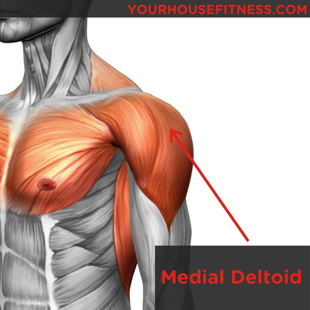
Muscle Breakdown Medial Deltoid

Deltoid Muscle Medical Art Library

The Deltoid Muscle Get the Basic Facts About It

Deltoid (Front, Lateral, Rear) Anatomy, Location, Function, Pain

Deltoid muscle anatomy, fibers, function and action of the deltoid muscle

VASTRAL PHYSIOTHERAPY CLINIC Deltoid Muscle Anatomy
Web Anatomy Where Are The Deltoid Muscles Located?
Your Assignment Is To Do Quicksketch Drawings Of The Deltoid From The Model Photos I’ve Provided In The Description Below.
Web Let’s Now Draw In The Shoulders On Top Of The Foundation In These Diagrams.
Web The Deltoid Muscle Is Known For Its Origin’s Distinctive ‘U’ Shape, Spanning Across The Clavicle, Acromion, And The Spine Of The Scapula.
Related Post: