Draw And Label A Microscope
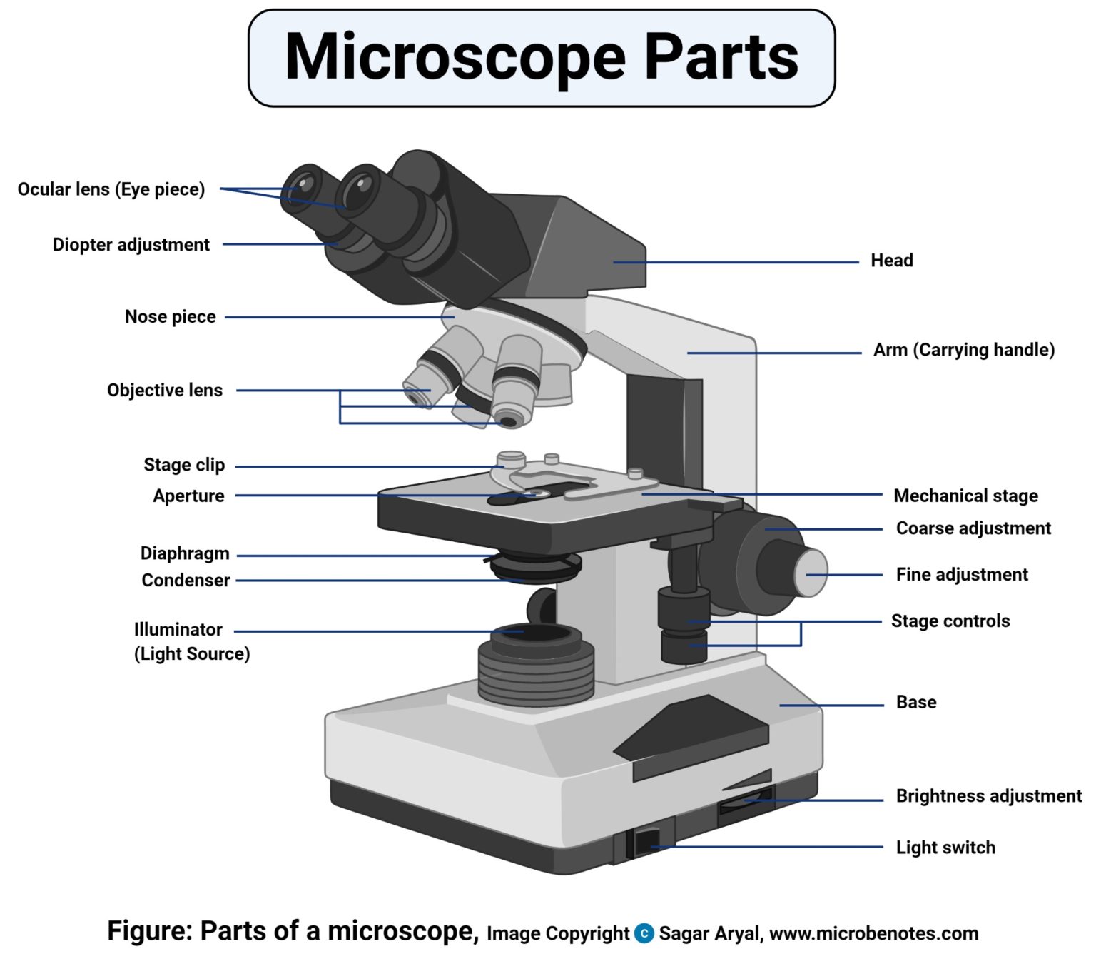
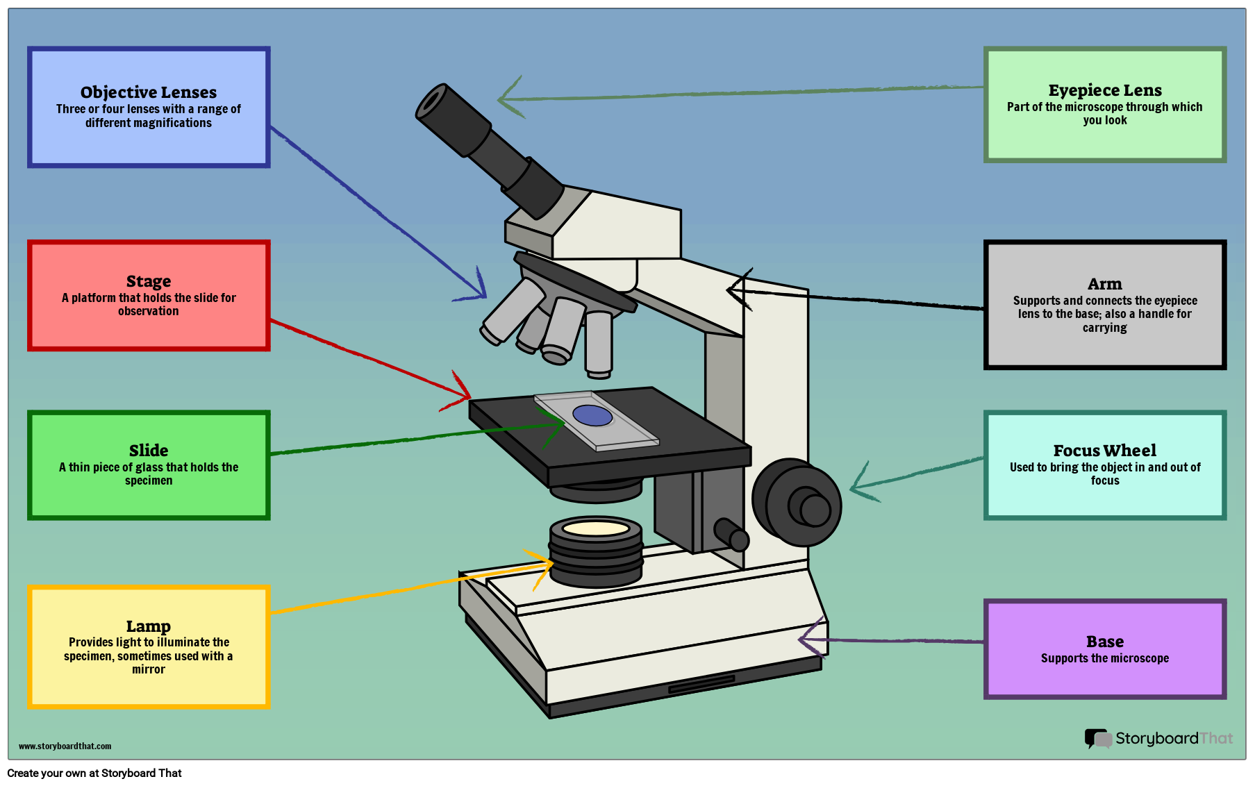
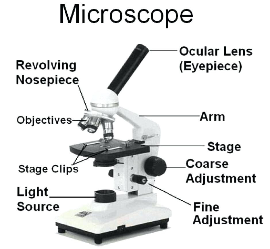
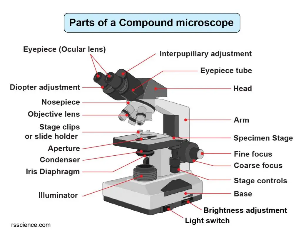
Draw And Label A Microscope - Draw the base of the microscope sketch 1.7 step 7: The eyepiece usually contains a 10x or 15x power lens. Useful as a study guide for learning the anatomy of a microscope. There are two major optical lens parts of a microscope: Be sure to indicate the magnification used and specimen name. Download the label the parts of the microscope pdf printable version here. Begin with the eyepiece 1.2 step 2: Outline the arm frame 1.4 step 4: Label the cell wall, cell membrane, cytoplasm, and chloroplasts in your lab manual. Web this short video discuss the expectations of a microscope observation and drawings and also provides examples of errors to watch out for.teachers: Web a microscope is one of the invaluable tools in the laboratory setting. The eyepiece usually contains a 10x or 15x power lens. However, as the saying goes, ‘practice makes perfect’, here is a blank compound microscope diagram. Young artists can learn to draw a microscope by following the photos in this easy guide. Kids will enjoy this simple step. Web this activity has been designed for use in homes and schools. In this tutorial, writing master shows you how to draw a realistic microscope with labels step by step. Web these labeled microscope diagrams and the functions of its various parts, attempt to simplify the microscope for you. Web simple microscope is a magnification apparatus that uses a combination. It is also called a body tube or eyepiece tube. Kids will enjoy this simple step by step lesson for learning how to draw this essential piece of scientific equipment. Diagram of parts of a microscope. Search for a diagram of a microscope. Stage and stage clips 7. Most photographs of cells are taken using a microscope, and these pictures can also be called micrographs. Perfect for students or anyone. Web each part of the compound microscope serves its own unique function, with each being important to the function of the scope as a whole. Kids will enjoy this simple step by step lesson for learning how to. It is used to observe things that cannot be seen by the naked eye. It is also called a body tube or eyepiece tube. Web simple microscope is a magnification apparatus that uses a combination of double convex lens to form an enlarged, erect image of a specimen. There are three structural parts of the microscope i.e. Useful as a. The eyepiece usually contains a 10x or 15x power lens. Useful as a study guide for learning the anatomy of a microscope. Web a microscope is an instrument that magnifies objects otherwise too small to be seen, producing an image in which the object appears larger. The body tube connects the eyepiece to the objective lenses. Also indicate the estimated. Each microscope layout (both blank and the version with answers) are available as pdf downloads. Web there are three major structural parts of a microscope: Be sure to indicate the magnification used and specimen name. Most photographs of cells are taken using a microscope, and these pictures can also be called micrographs. Web simple microscope is a magnification apparatus that. Continue follow my channel and like, share,comment also. There are two major optical lens parts of a microscope: Use a landscape poster layout (large or small). Diagram of parts of a microscope. Web a microscope is an instrument that magnifies objects otherwise too small to be seen, producing an image in which the object appears larger. Web these labeled microscope diagrams and the functions of its various parts, attempt to simplify the microscope for you. The individual parts of a compound microscope can vary heavily depending on the configuration & applications that the scope is being used for. To facilitate the scrutiny of such miniature entities with unparalleled. Useful as a study guide for learning the. The body tube connects the eyepiece to the objective lenses. Perfect for students or anyone. The working principle of a simple microscope is that when a lens is held close to the eye, a virtual, magnified and erect image of a specimen is formed at the least possible distance from which a human. The lens the viewer looks through to. The scientific discipline encompassing the study of these diminutive entities through a microscope is called microscopy. Web how to draw a microscope 🔬. The body tube connects the eyepiece to the objective lenses. Web these labeled microscope diagrams and the functions of its various parts, attempt to simplify the microscope for you. Perfect for students or anyone. First and foremost, we have a labeled microscope diagram, available in both black and white and color. There are two major optical lens parts of a microscope: Continue follow my channel and like, share,comment also. Outline the slide platform 1.6 step 6: Draw the base of the microscope sketch 1.7 step 7: Draw the objective lenses 1.5 step 5: Web there are three major structural parts of a microscope: Be sure to indicate the magnification used and specimen name. Web in this interactive, you can label the different parts of a microscope. Eyepiece (10x) and objective lenses (4x, 10x, 40x, 100x). Useful as a study guide for learning the anatomy of a microscope.
Parts of a microscope with functions and labeled diagram

5 Types of Microscopes with Definitions, Principle, Uses, Labeled Diagrams

Microscope diagram Tom Butler Science skills, Microscope parts

Microscope Drawing And Label at GetDrawings Free download

36+ Label Each Part Of A Microscope Gif Diagram Printabel

Microscope Diagram Labeled, Unlabeled and Blank Parts of a Microscope

How to Use a Microscope (Properly) Step by Step New York Microscope

Labeled Microscope Diagram Tim's Printables

Parts Of A Microscope With Functions And Labeled Diagram Images

Compound Microscope Parts Labeled Diagram and their Functions (2023)
Web A Microscope Is A Laboratory Instrument Employed To Examine Minute Entities, Such As Cells And Microorganisms, Which Are Invisible To The Naked Eye.
Outline The Arm Frame 1.4 Step 4:
Also Indicate The Estimated Cell Size In Micrometers Under Your Drawing.
Always Lift A Microscope By Holding Both The Arm And Base With Two Hands.
Related Post: