Drawing Spinal Cord
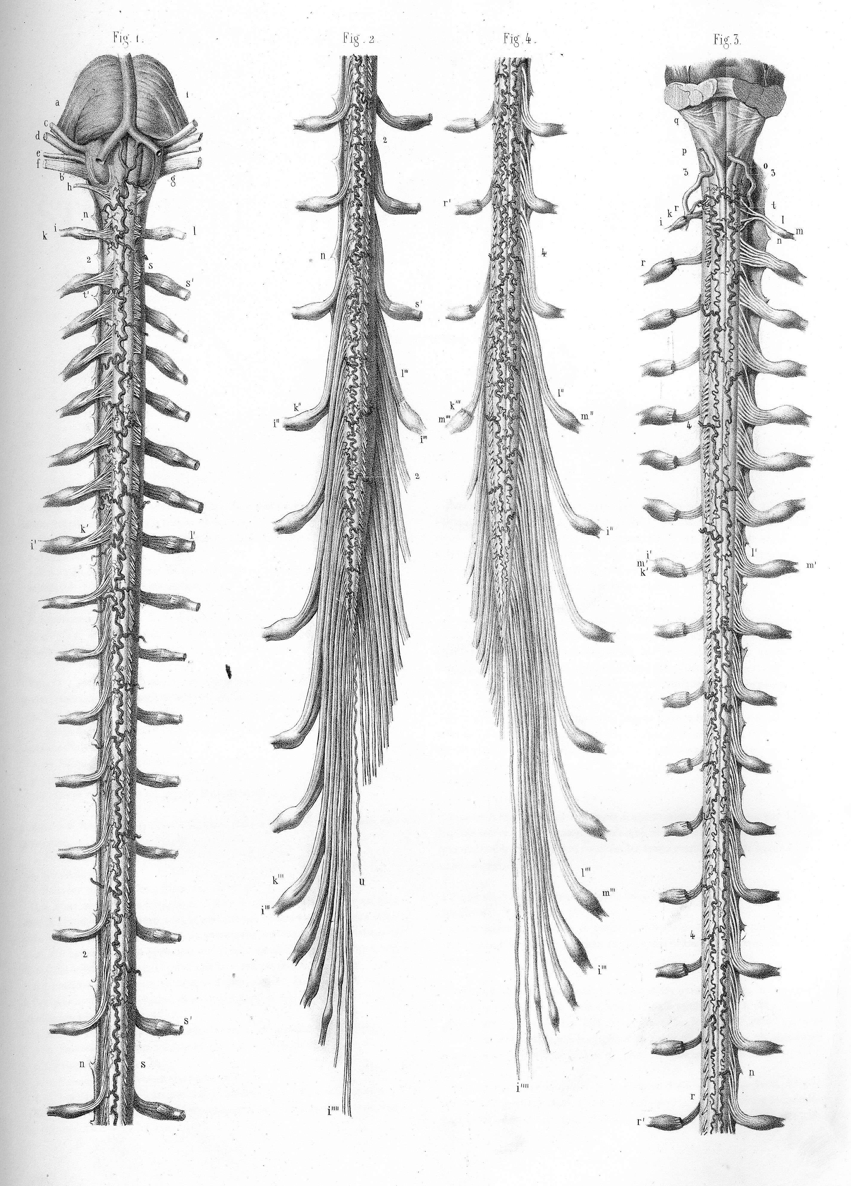
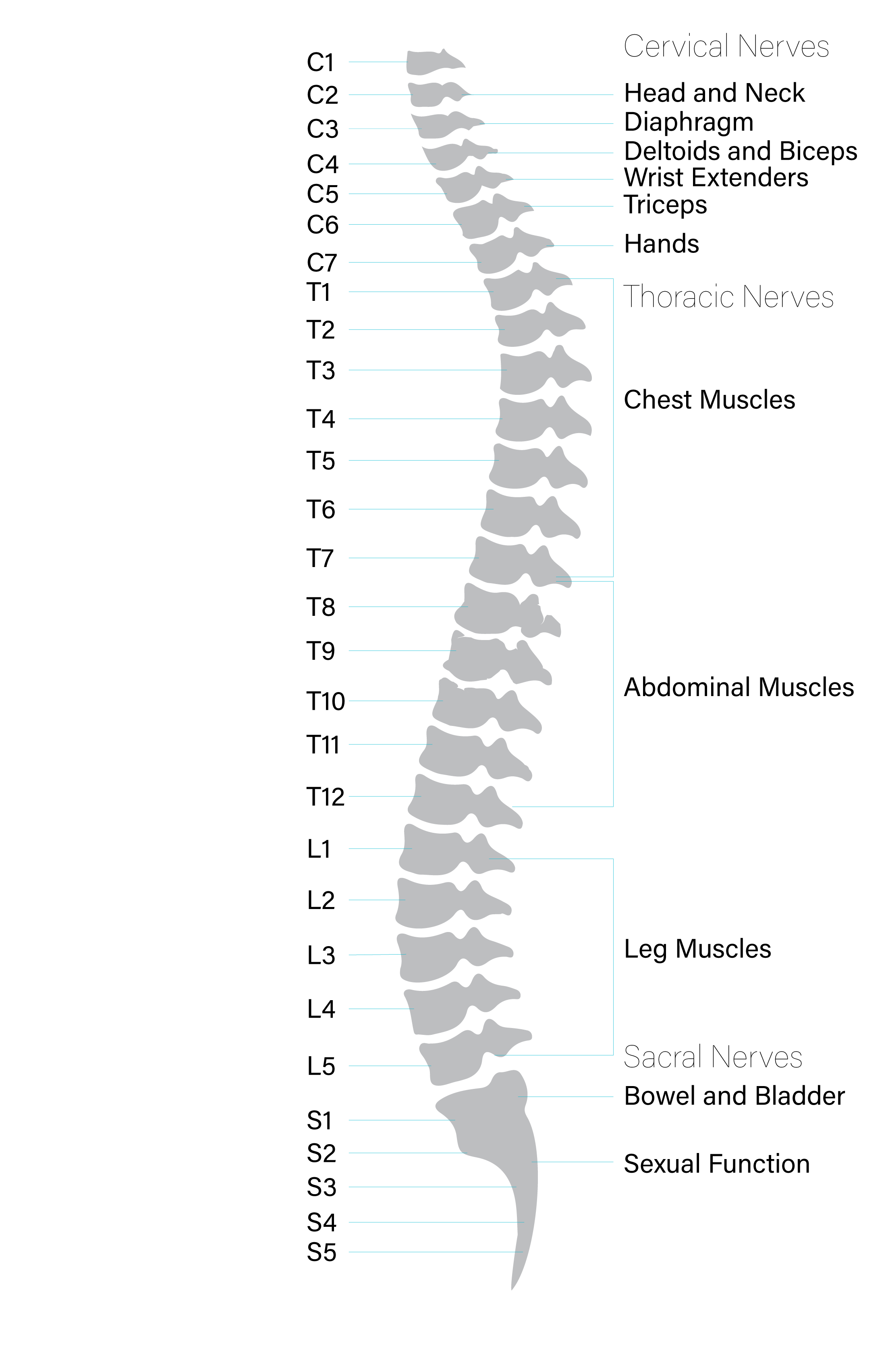
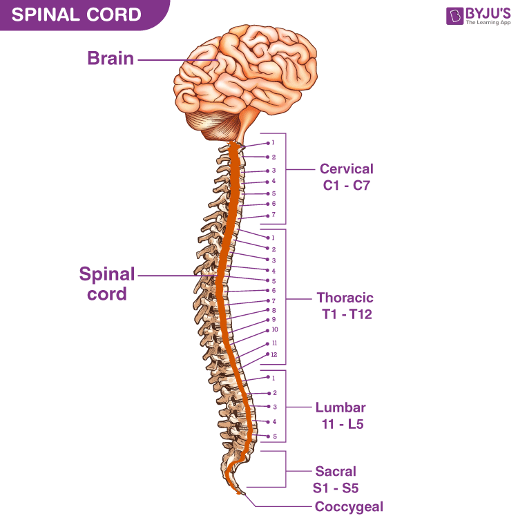
Drawing Spinal Cord - It was designed particularly for physiotherapists, osteopaths, rheumatologists, neurosurgeons, orthopedic surgeons and general practitioners, especially for the study and understanding of medullary diseases. Your spinal cord helps carry electrical nerve signals throughout your body. Next, add in the details. Web how to draw human spinal cord step by stepin this video, i share how to draw step by step using basic drawing techniques. Web the spinal cord is part of the central nervous system (cns), which extends caudally and is protected by the bony structures of the vertebral column. These nerve signals help you feel sensations and move your muscles. It contains tissues, fluids and nerve cells. Web this atlas of human anatomy describes the spinal cord through 18 anatomical diagrams with 270 anatomical structures labeled. Web the spinal cord is a tubular bundle of nervous tissue and supporting cells that extends from the brainstem to the lumbar vertebrae. According to some estimates, females have a spinal cord of about 43 centimeters (cm), while males have a spinal cord. Also available for free download. The cerebrum, the diencephalon, the brain stem, and the cerebellum. Web the trial involved 20 patients, with half receiving active spinal cord stimulation and the other half a control version. Cervical, thoracic, lumbar and sacral. Web 3 views 5 days ago. This includes adding in the ridges on the spinal cord, as well as the tunnels that run down the center. Web each segment of the spinal cord provides several pairs of spinal nerves, which exit from. Web © 2023 google llc hi, viewers! Sacral cord lumbar cord thoracic cord cervical cord coccygeal The cerebrum, the diencephalon, the brain stem, and. Web open access june 8, 2017 teaching spinal cord neuroanatomy through drawing: In this video i will guide you through the creation of your own drawing of a spinal cord cross section.i am creating these videos to further supplement the v. Diagram of the spinal cord is illustrated in detail with neat and clear labelling. Web explore spinal cord diagram. A person’s conscious experiences are based on neural activity in the brain. It was designed particularly for physiotherapists, osteopaths, rheumatologists, neurosurgeons, orthopedic surgeons and general practitioners, especially for the study and understanding of medullary diseases. Web basics terminologyintroduction to the musculoskeletal systemintroduction to the other systems upper limb overviewshoulder and armelbow and forearmwrist and handnerves and vessels lower limb overviewhip. The spinal cord is divided into five different parts. Web open access june 8, 2017 teaching spinal cord neuroanatomy through drawing: Of spinal cord (peripheral) giribabu biology 9.67k subscribers subscribe 78k views 3 years ago spinal cord transevers section diagram to. Web this atlas of human anatomy describes the spinal cord through 18 anatomical diagrams with 270 anatomical structures labeled.. The spinal cord is divided into five different parts. Web your spinal cord is the long, cylindrical structure that connects your brain and lower back. Web © 2023 google llc hi, viewers! It forms a vital link between the brain and the body. Web how to draw human spinal cord step by stepin this video, i share how to draw. Web the spinal cord is a single structure, whereas the adult brain is described in terms of four major regions: Web each segment of the spinal cord provides several pairs of spinal nerves, which exit from. Web 5 years ago the olfactory nerve (1st), the optic nerve (2nd), oculomotor nerve (3rd), trochlear nerve (4th), trigeminal nerve (5th), abducens nerve (6th),. Web your spinal cord is the long, cylindrical structure that connects your brain and lower back. Next, add in the details. Designated space for drawing is provided and labeled on the associated answer packet ( appendix b ). Web basics terminologyintroduction to the musculoskeletal systemintroduction to the other systems upper limb overviewshoulder and armelbow and forearmwrist and handnerves and vessels. Web anatomy structure injuries nerves functions spinal cord anatomy in adults, the spinal cord is usually 40cm long and 2cm wide. Many of the nerves of the peripheral nervous system, or pns, branch out from the spinal cord and travel to. Web each segment of the spinal cord provides several pairs of spinal nerves, which exit from. It forms a. The cerebrum, the diencephalon, the brain stem, and the cerebellum. The regulation of homeostasis is governed by a specialized region in the brain. Web anatomy the length of the spinal cord varies from person to person. The sacral region has a tapered end called the conus medullaris. Web figure 12.6.1 12.6. A person’s conscious experiences are based on neural activity in the brain. Web anatomy structure injuries nerves functions spinal cord anatomy in adults, the spinal cord is usually 40cm long and 2cm wide. Of spinal cord (peripheral) giribabu biology 9.67k subscribers subscribe 78k views 3 years ago spinal cord transevers section diagram to. Web the trial involved 20 patients, with half receiving active spinal cord stimulation and the other half a control version. The regulation of homeostasis is governed by a specialized region in the brain. Web figure 12.6.1 12.6. Web the spinal cord is a single structure, whereas the adult brain is described in terms of four major regions: Web the following is a general guide on how to draw a spinal cord. The sacral region has a tapered end called the conus medullaris. Web the spinal cord begins at the base of the brain and extends into the pelvis. Web 5 years ago the olfactory nerve (1st), the optic nerve (2nd), oculomotor nerve (3rd), trochlear nerve (4th), trigeminal nerve (5th), abducens nerve (6th), facial nerve (7th), vestibulocochlear nerve (8th), glossopharyngeal nerve (9th), vagus nerve (10th), accessory nerve (1th), and hypoglossal nerve (12th) 1 comment ( 42 votes) upvote Web this atlas of human anatomy describes the spinal cord through 18 anatomical diagrams with 270 anatomical structures labeled. These nerve signals help you feel sensations and move your muscles. In this video i will guide you through the creation of your own drawing of a spinal cord cross section.i am creating these videos to further supplement the v. Next, add in the details. It is covered by the three membranes of the cns, i.e., the dura mater, arachnoid and the innermost pia mater.
Spinal Cord Drawing at Explore collection of

How Does The Spinal Cord Work Reeve Foundation

Spinal Cord Drawing at GetDrawings Free download

spinal cord Spine drawing, Anatomy art, Skeleton drawings

Spinal Cord, Drawing Stock Image C017/1520 Science Photo Library

Anatomy of the spinal cord. Download Scientific Diagram

The structure and segments of the spinal cord Medical diagnosis

The Spinal Cord Neurologic Clinics

Anatomy of the Spinal Cord Praxis Spinal Cord Institute

Spinal Cord Anatomy, Structure, Function, & Diagram
Sacral Cord Lumbar Cord Thoracic Cord Cervical Cord Coccygeal
In This Section, Students Are Instructed To Draw Out Various Components Of Spinal Cord Anatomy.
How To Draw Structure Of The Spinal Cord Diagram.
It Forms A Vital Link Between The Brain And The Body.
Related Post: