Gram Positive Chart
Gram Positive Chart - Identify similarities and differences between high g+c and low g+c bacterial groups. Web after your bacteria have been isolated and you have good results from your gram stain, begin to follow the flow chart below. Continuing education (ce) this course does not offer p.a.c.e ® credits. Identify similarities and differences between high g+c and low g+c bacterial groups. These categories are based on their cell wall composition and reaction to the gram stain test. But, some bacteria stain either as gram positive or gram negative, depending on conditions. Most bacteria are classified into two broad categories: Interpret the results of biochemical methods. Microscopically, staphylococci appear in grapelike clusters, whereas streptococci are in chains. In a gram stain test, these organisms yield a positive result. Web gram positive bacteria types and classification. Give an example of a bacterium of high g+c and low g+c group commonly associated with each category. Gram project is a medical education resource website containing diagrams, tables and flowcharts for all your quick referencing, revision and teaching needs. Web updated on february 05, 2020. 1 interpretation of key phrases. Web gram positive bacteria types and classification. Web gram positive bacteria have a thick coating of peptidoglycan and stain purple with crystal violet. Here’s why knowing whether the result is positive or negative is. Microscopically, staphylococci appear in grapelike clusters, whereas streptococci are in chains. Interpret the results of biochemical methods. Continuing education (ce) this course does not offer p.a.c.e ® credits. Give an example of a bacterium of high g+c and low g+c group commonly associated with each category. Hans christian gram developed the staining method in 1884. Here’s why knowing whether the result is positive or negative is. Web gram positive bacteria have a thick coating of peptidoglycan and. Give an example of a bacterium of high g+c and low g+c group commonly associated with each category. These categories are based on their cell wall composition and reaction to the gram stain test. Actinomyces, bacillus, clostridium, corynebacterium, enterococcus, gardnerella, lactobacillus, listeria, mycoplasma, nocardia, staphylococcus, streptococcus, streptomyces ,etc. Gram positive and gram negative. Here’s why knowing whether the result is. Hans christian gram developed the staining method in 1884. In a gram stain test, these organisms yield a positive result. Web updated on february 05, 2020. (redirected from gram positive bacteria) contents. Web after your bacteria have been isolated and you have good results from your gram stain, begin to follow the flow chart below. Web updated on february 05, 2020. In a gram stain test, these organisms yield a positive result. During the gram staining process — a test that experts use to view the bacteria under a microscope — they appear purple or. Gram positive and gram negative. But, some bacteria stain either as gram positive or gram negative, depending on conditions. Web use flowcharts and identification charts to identify some common aerobic gram positive microorganisms. Web gram positive bacteria have a thick coating of peptidoglycan and stain purple with crystal violet. Interpret the results of biochemical methods. Most bacteria are classified into two broad categories: Actinomyces, bacillus, clostridium, corynebacterium, enterococcus, gardnerella, lactobacillus, listeria, mycoplasma, nocardia, staphylococcus, streptococcus, streptomyces ,etc. Microscopically, staphylococci appear in grapelike clusters, whereas streptococci are in chains. Web aerobic gram positive rods flowchart. But, some bacteria stain either as gram positive or gram negative, depending on conditions. Gram positive and gram negative. Give an example of a bacterium of high g+c and low g+c group commonly associated with each category. Web use flowcharts and identification charts to identify some common aerobic gram positive microorganisms. (redirected from gram positive bacteria) contents. They don’t retain crystal violet, so are stained red or pink with carbol fuchsin or safranin. In a gram stain test, these organisms yield a positive result. These categories are based on their cell wall composition and reaction to the. Actinomyces, bacillus, clostridium, corynebacterium, enterococcus, gardnerella, lactobacillus, listeria, mycoplasma, nocardia, staphylococcus, streptococcus, streptomyces ,etc. They don’t retain crystal violet, so are stained red or pink with carbol fuchsin or safranin. Hans christian gram developed the staining method in 1884. Web gram positive bacteria have a thick coating of peptidoglycan and stain purple with crystal violet. Identify similarities and differences between. Identify similarities and differences between high g+c and low g+c bacterial groups. Most bacteria are classified into two broad categories: Gram project is a medical education resource website containing diagrams, tables and flowcharts for all your quick referencing, revision and teaching needs. (redirected from gram positive bacteria) contents. 1 interpretation of key phrases. Here’s why knowing whether the result is positive or negative is. Hans christian gram developed the staining method in 1884. Associate various biochemical tests with their correct applications. But, some bacteria stain either as gram positive or gram negative, depending on conditions. Gram negative bacteria lack this thick coating. Microscopically, staphylococci appear in grapelike clusters, whereas streptococci are in chains. Give an example of a bacterium of high g+c and low g+c group commonly associated with each category. Web updated on february 05, 2020. They don’t retain crystal violet, so are stained red or pink with carbol fuchsin or safranin. The bacterial cell wall of these organisms have thick peptidoglycan layers, which take up the purple/violet stain. Web aerobic gram positive rods flowchart.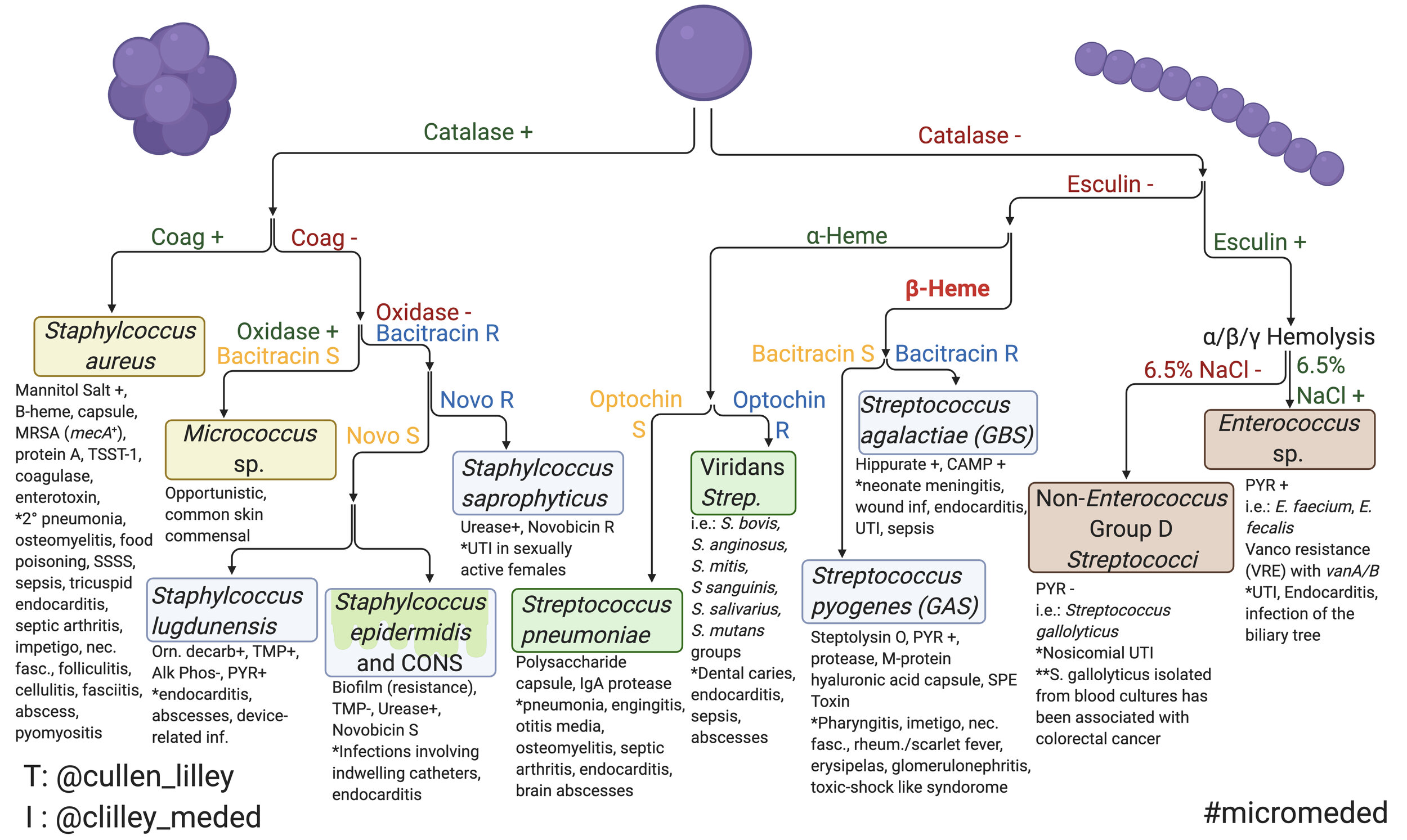
Gram Negative Cocci Flow Chart

MBBS Medicine (Humanity First) February 2014

Pin by Rachel Noble on MICROBIOLOGY rotation Pinterest
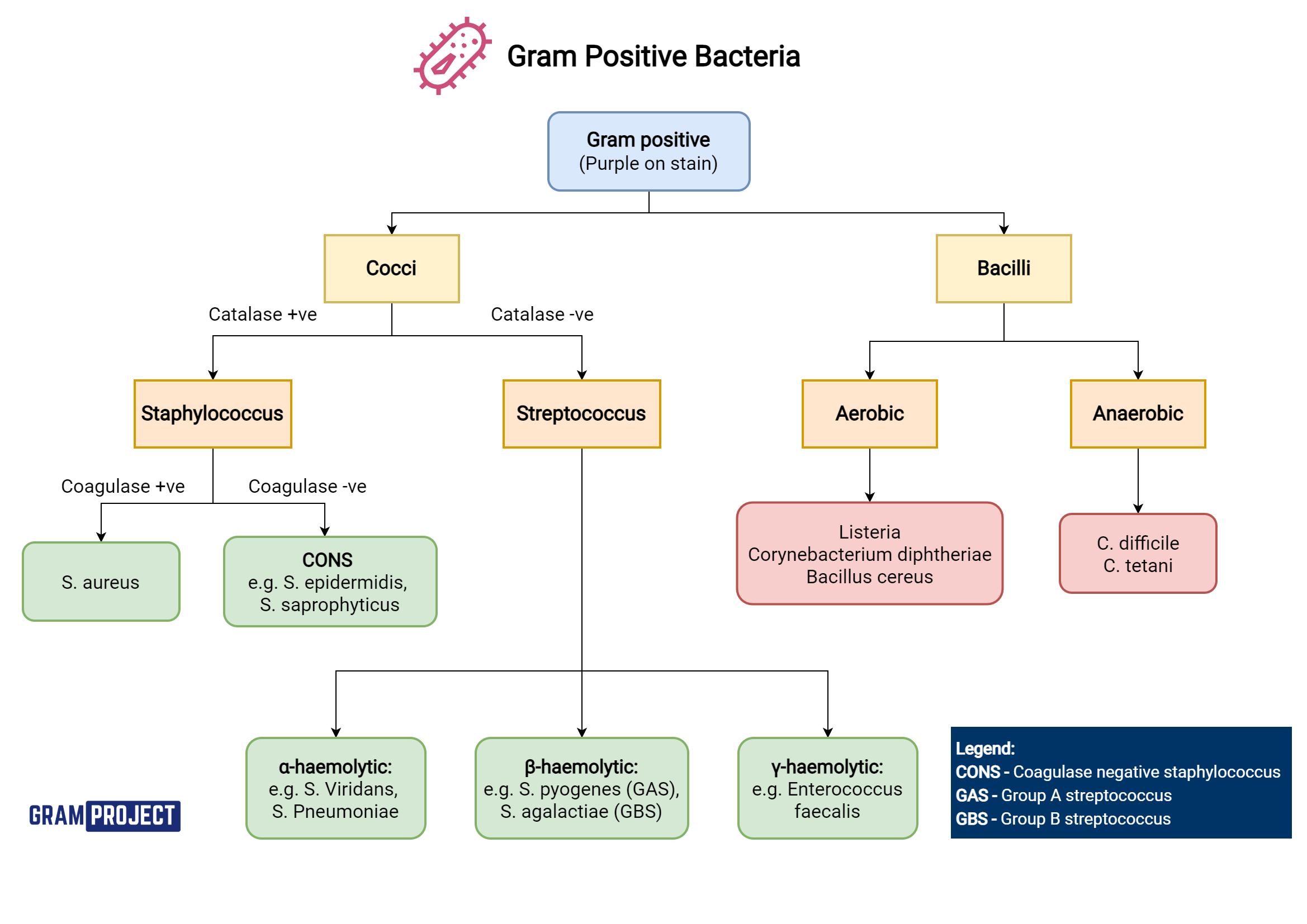
Gram Positive Organisms Chart

Gram () Chart Diagram Quizlet
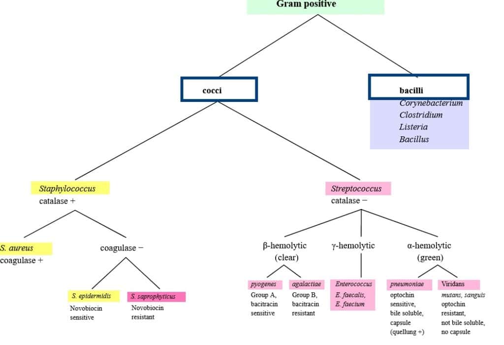
Gram Positive Organisms Chart

Gram Positive Bacteria Overview Identification Algorithm GrepMed

Gram Negative Cocci Flow Chart

Gram Positive Organisms Chart

Streptococcus Concise Medical Knowledge
Gram Positive And Gram Negative.
Web Use Flowcharts And Identification Charts To Identify Some Common Aerobic Gram Positive Microorganisms.
These Categories Are Based On Their Cell Wall Composition And Reaction To The Gram Stain Test.
During The Gram Staining Process — A Test That Experts Use To View The Bacteria Under A Microscope — They Appear Purple Or.
Related Post: