Mri Tarlov Cyst Size Chart
Mri Tarlov Cyst Size Chart - Web tarlov’s cysts on mri. Your physician might order an mri and then refer you to a specialist—such as a neurosurgeon—if the mri shows a cyst. Tarlov’s cysts appear white in t2 images. Water densities have a high‐intensity signal (with white appearance) on t2 weighted mri. Signal characteristics of typical tarlov cyst are those of csf on all sequences. Web tarlov cysts vary in size. Web tarlov cysts may be discovered when patients with low back pain or sciatica have a magnetic resonance imaging (mri) performed. Web most tarlov cysts are discovered on mri, ct or myelogram. Web figures 1,2 1,2 demonstrate lumbosacral mri for symptomatic tarlov’s cyst before and after fenestration, respectively. You might hear your healthcare provider refer to tarlov cysts as meningeal cysts or perineural cysts. On mr the perineural cysts predominantly originating near the sacral nerves adjacent to the dorsal root ganglion. If you’re experiencing symptoms of tarlov cysts or back pain in general, talk with your primary care physician. The cysts appear in the roots of the nerves that grow out of the spinal cord. Web the sacral foramina may be widened. Web tarlov. Web mri of giant multiple tarlov cysts. Web tarlov cysts vary in size. Shock or trauma of the spine, or exertion, can cause spinal fluid in the cysts to build up. Your physician might order an mri and then refer you to a specialist—such as a neurosurgeon—if the mri shows a cyst. On mr the perineural cysts predominantly originating near. Your physician might order an mri and then refer you to a specialist—such as a neurosurgeon—if the mri shows a cyst. Web mri, or magnetic resonance imaging, is considered the imaging study of choice in identifying tarlov cysts. Web tarlov cysts vary in size. Web tarlov cysts may be discovered when patients with low back pain or sciatica have a. Multiformat reformation (mpr) from 3 dimensional t1 mri sequence. Mri provides better resolution of tissue density, absence of bone interference, multiplanar capabilities, and is noninvasive. Tarlov’s cysts appear white in t2 images. Individuals may be affected by multiple cysts of varying size. You might hear your healthcare provider refer to tarlov cysts as meningeal cysts or perineural cysts. Larger cysts may cause symptoms like pain, bowel and bladder problems or numbness. On mr the perineural cysts predominantly originating near the sacral nerves adjacent to the dorsal root ganglion. Symptoms can occur depending upon the size and specific location of. Web the sacral foramina may be widened. When not documented, a study investigator reviewed mri images. Tarlov’s cysts appear white in t2 images. Plain films may show bony erosion of the spinal canal or of the sacral foramina. Mri revealed 8 tarlov cysts (size: Most cysts are completely asymptomatic and are found incidentally when working up a different spinal issue. Web the sacral foramina may be widened. Web most tarlov cysts are discovered on mri, ct or myelogram. On mr the perineural cysts predominantly originating near the sacral nerves adjacent to the dorsal root ganglion. Symptoms can occur depending upon the size and specific location of. Shock or trauma of the spine, or exertion, can cause spinal fluid in the cysts to build up. Axial ( a. Shock or trauma of the spine, or exertion, can cause spinal fluid in the cysts to build up. If you’re experiencing symptoms of tarlov cysts or back pain in general, talk with your primary care physician. Web most tarlov cysts are discovered on mri, ct or myelogram. The widest diameter of each tarlov cyst in the sagittal plane was documented. It is sometimes confusing to make an accurate diagnosis as to the cause of the symptoms, if there are multiple diagnoses found, such as herniated discs , ruptured disc or degenerative disc disease (. Mri revealed 8 tarlov cysts (size: Your physician might order an mri and then refer you to a specialist—such as a neurosurgeon—if the mri shows a. Web tarlov cyst size and location were abstracted from the radiology report when present; Retrospective chart review was conducted for 220 patients with tarlov cysts seen at our institution between 2006 and 2021. Morphology can vary from a simple rounded cyst to a complex loculated cystic mass with septations. Most cysts are completely asymptomatic and are found incidentally when working. Most cysts are completely asymptomatic and are found incidentally when working up a different spinal issue. Morphology can vary from a simple rounded cyst to a complex loculated cystic mass with septations. Web mri, or magnetic resonance imaging, is considered the imaging study of choice in identifying tarlov cysts. Web tarlov cysts were most commonly located from s2 to s3 (73%), and ranged in size from 1 to 2 cm (55%). Mri revealed 8 tarlov cysts (size: Larger cysts may cause symptoms like pain, bowel and bladder problems or numbness. If you’re experiencing symptoms of tarlov cysts or back pain in general, talk with your primary care physician. Symptoms can occur depending upon the size and specific location of. Web figures 1,2 1,2 demonstrate lumbosacral mri for symptomatic tarlov’s cyst before and after fenestration, respectively. Retrospective chart review was conducted for 220 patients with tarlov cysts seen at our institution between 2006 and 2021. Web tarlov cysts vary in size. Web most tarlov cysts are discovered on mri, ct or myelogram. We prefer a closed mri without contrast/dye. Web most tarlov cysts are discovered on mri, ct or myelogram. This sagital view of a t2 weighted mri reveals a hyper‐intense signal posterior to the s2 segment. Shock or trauma of the spine, or exertion, can cause spinal fluid in the cysts to build up.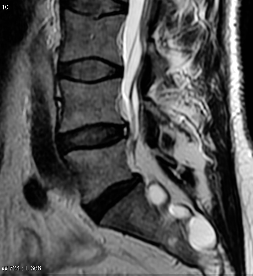
Tarlov Cysts WikiMSK
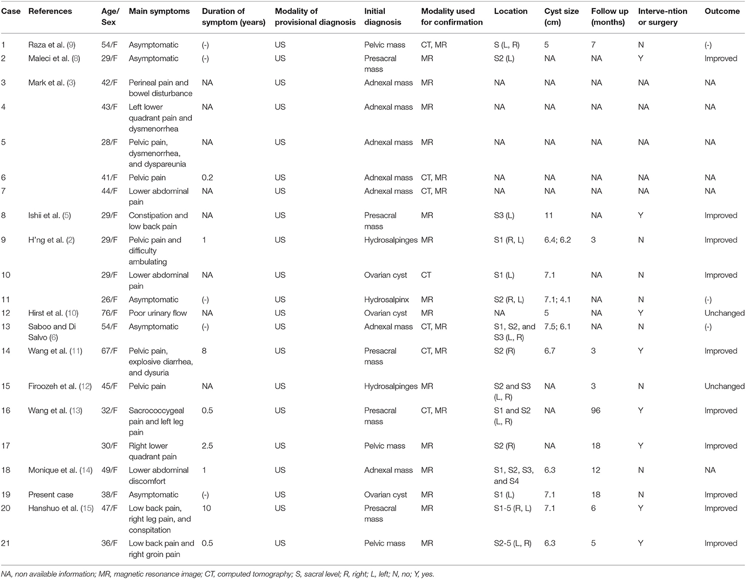
Tarlov Cyst Size Chart
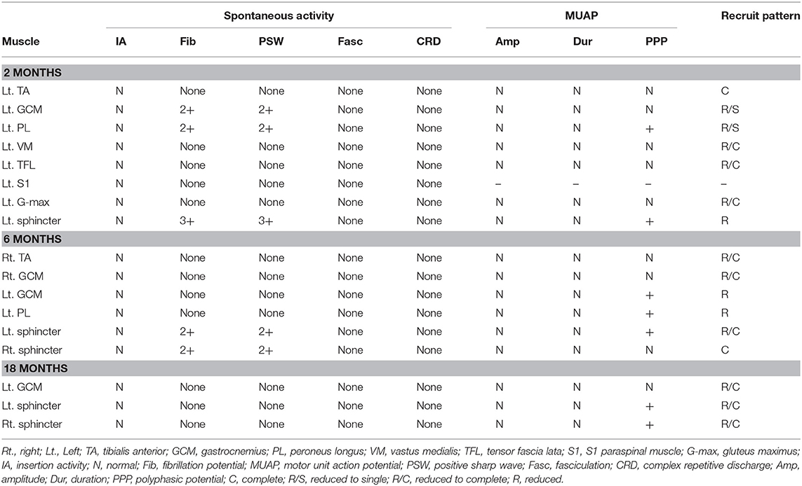
Tarlov Cyst Size Chart

Tarlov Cyst Excel Spine

Tarlov Cyst Size Chart

Tarlov Cyst Size Chart
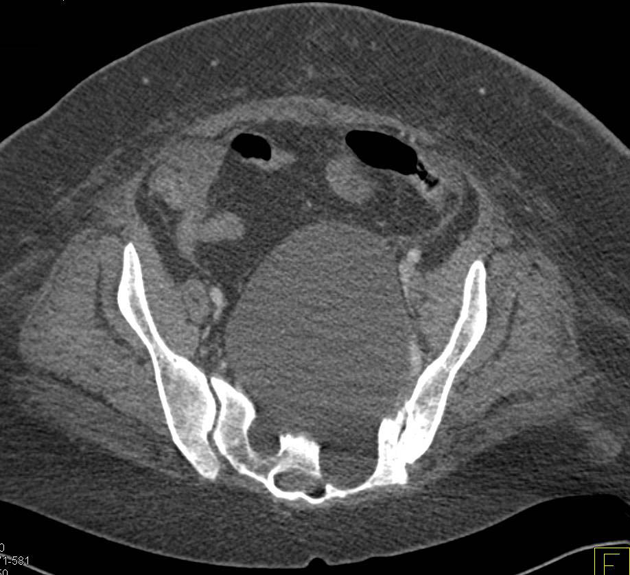
Tarlov Cyst Size Chart
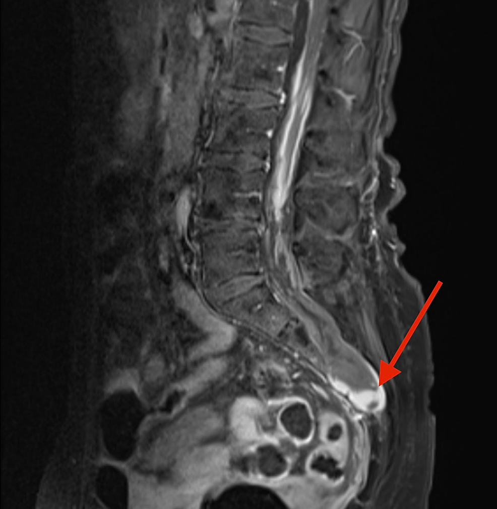
Cureus Tarlov Cyst Rupture and Intradural Hemorrhage Mimicking

(PDF) Tarlov Cysts A Report of Two Cases

Treatment of 213 Patients with Symptomatic Tarlov Cysts by CTGuided
Figures 3,4 3,4 Demonstrate Surgical Operation For Symptomatic Tarlov’s Cyst(S) With Nerve Root Imbrication.
You Might Hear Your Healthcare Provider Refer To Tarlov Cysts As Meningeal Cysts Or Perineural Cysts.
Web Tarlov Cysts May Be Discovered When Patients With Low Back Pain Or Sciatica Have A Magnetic Resonance Imaging (Mri) Performed.
Web Tarlov’s Cysts On Mri.
Related Post: