Nerves Of The Spine Chart
Nerves Of The Spine Chart - Web there are 31 pairs of spinal nerves. On the chart below you will see 4 columns (vertebral level, nerve root, innervation, and possible symptoms). Thoracic spinal nerves are not part of any plexus, but give rise to the intercostal nerves directly. Web what does the spine do? Each of these nerves branches out from the spinal cord, dividing and subdividing to form a network connecting the spinal cord to every part of the body. Type 1 neurofibromatosis (nf1) is the most common neurocutaneous disorder, and it is an inherited condition that causes a tumour predisposition. Central nervous system (cns) manifestations are a significant cause of morbidity and mortality in nf1. Web the spinal cord and peripheral nerves. This root has a swelling called the dorsal root ganglion, which contains the cell bodies of sensory neurons. Each pair connects the spinal cord with a specific region of the body. The peripheral nerves are responsible for sensations and muscle movements. For most spinal segments, the nerve roots run through the bony canal, and at each level a pair of nerve roots exits from the spine. The back functions are many, such as to house and protect the spinal cord, hold the body and head upright, and adjust the movements of. Web spinal nerves are all mixed nerves with both sensory and motor fibers. Each spinal nerve is attached to the spinal cord by two roots: Each spinal nerve originates from two roots: The spinal cord transmits signals from the brain to the rest of a person’s body. Web there are 5 pairs of lumbar spinal nerves that progressively increase in. Web learn the anatomy of the spinal nerves, including their roots, components and functions faster and more efficiently with this comprehensive article. Spinal nerves emerge from the spinal cord and reorganize through plexuses, which then give rise to systemic nerves. Each spinal nerve originates from two roots: Protecting your spinal cord (nerves that connect your brain to the rest of. A dorsal (or posterior) root which relays sensory information and a ventral (or anterior) root which relays motor information. We provide a pictorial review of neuroradiological. Paul slosar, md, orthopedic surgeon. In total, there are 31 pairs of spinal nerves grouped regionally by spinal region. These complex networks of nerves enable the brain to receive sensory inputs from the skin. On the chart below you will see 4 columns (vertebral level, nerve root, innervation, and possible symptoms). Web to understand this intricate region, we will consider the bony structures first, and then discuss the ligaments, nerves, and musculature that are associated with this region of the spinal column, concluding with some clinical implications of damage to some of these structures.. Carries sensory (afferent) information to the spinal cord. These nerves exit the intervertebral foramina below the corresponding vertebra. Protecting your spinal cord (nerves that connect your brain to the rest of your body). Each spinal nerve is attached to the spinal cord by two roots: Giving your body structure (shape). In the neck, the nerve root is named for the lower segment that it runs between (e.g. Web each pair of spinal nerves roughly correspond to a segment of the vertebral column: Carries sensory (afferent) information to the spinal cord. Web the spinal cord and peripheral nerves. Again, they are named according to where they each exit in the spine. Each of these nerves branches out from the spinal cord, dividing and subdividing to form a network connecting the spinal cord to every part of the body. Web there are 31 pairs of spinal nerves. The point at which a nerve exits the spinal cord is called a nerve root. 8 cervical, 12 thoracic, 5 lumbar, 5 sacral, and 1. Allowing you to be flexible and move. Where is the spine located? Web the arthritis foundation states that 31 pairs of nerves branch off the spinal cord to other parts of the body. Web 5 sacral spinal nerves. This article will explore the key factors that impact visualizing spinal nerves and the importance of considering these factors when making decisions. Giving your body structure (shape). Each pair connects the spinal cord with a specific region of the body. Web spinal nerves are all mixed nerves with both sensory and motor fibers. The spinal cord transmits signals from the brain to the rest of a person’s body. Eight cervical spinal nerve pairs, 12 thoracic pairs , five lumbar pairs, five sacral. Type 1 neurofibromatosis (nf1) is the most common neurocutaneous disorder, and it is an inherited condition that causes a tumour predisposition. Protecting your spinal cord (nerves that connect your brain to the rest of your body). Web to understand this intricate region, we will consider the bony structures first, and then discuss the ligaments, nerves, and musculature that are associated with this region of the spinal column, concluding with some clinical implications of damage to some of these structures. The vertebral column’s most important physiologic function is protecting the spinal cord, which is the main avenue for communication between the brain and the. Eight cervical spinal nerves on each side of the spine called c1 through c8. Spinal nerves emerge from the spinal cord and reorganize through plexuses, which then give rise to systemic nerves. On the chart below you will see 4 columns (vertebral level, nerve root, innervation, and possible symptoms). Web spinal nerves are all mixed nerves with both sensory and motor fibers. Web there are 31 pairs of spinal nerves: This root has a swelling called the dorsal root ganglion, which contains the cell bodies of sensory neurons. Allowing you to be flexible and move. Again, they are named according to where they each exit in the spine (see figure below). It comprises the vertebral column (spine) and two compartments of back muscles; The point at which a nerve exits the spinal cord is called a nerve root. Giving your body structure (shape). Web there are 5 pairs of lumbar spinal nerves that progressively increase in size from l1 to l5.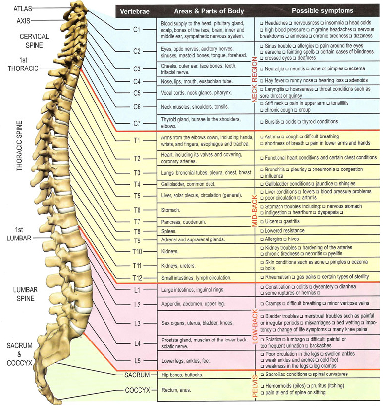
spine and nerve chart
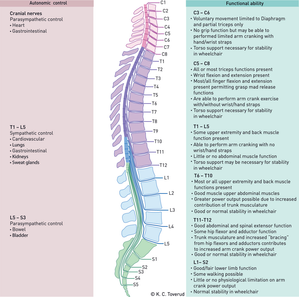
Anatomy Chart Spinal Nerves

Spinal Nerve Function Anatomical Chart Anatomy Models and Anatomical
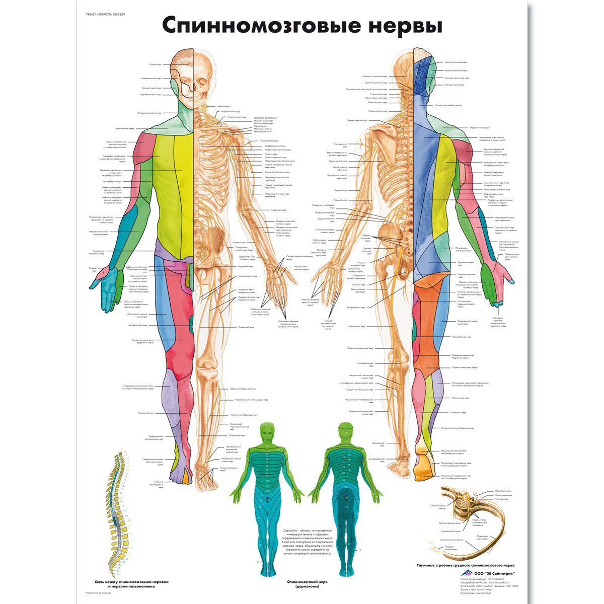
Spinal Nerves Chart 1002329 VR6621L 3B Scientific ZVR6621L
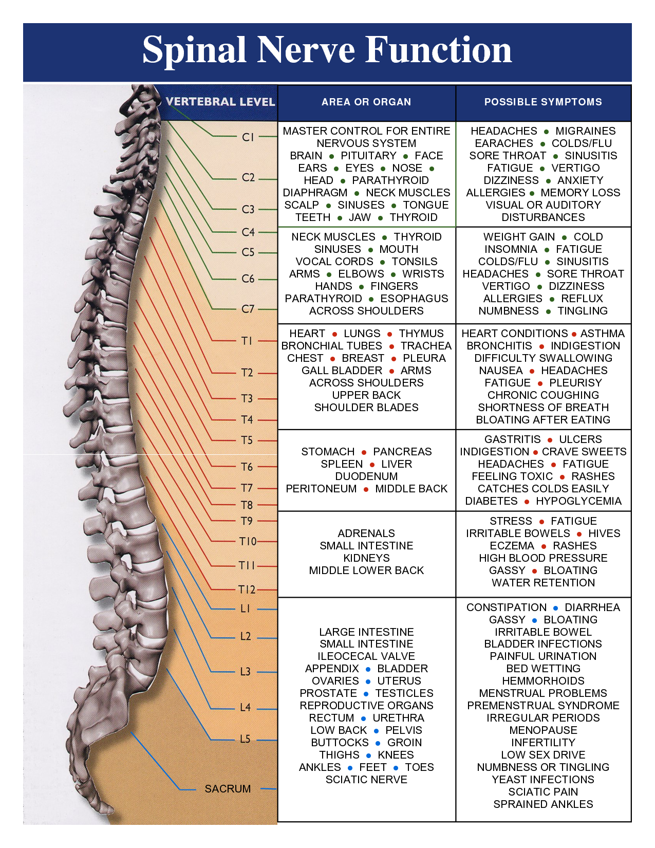
Vital Connections Briggs Chiropractic Clinic
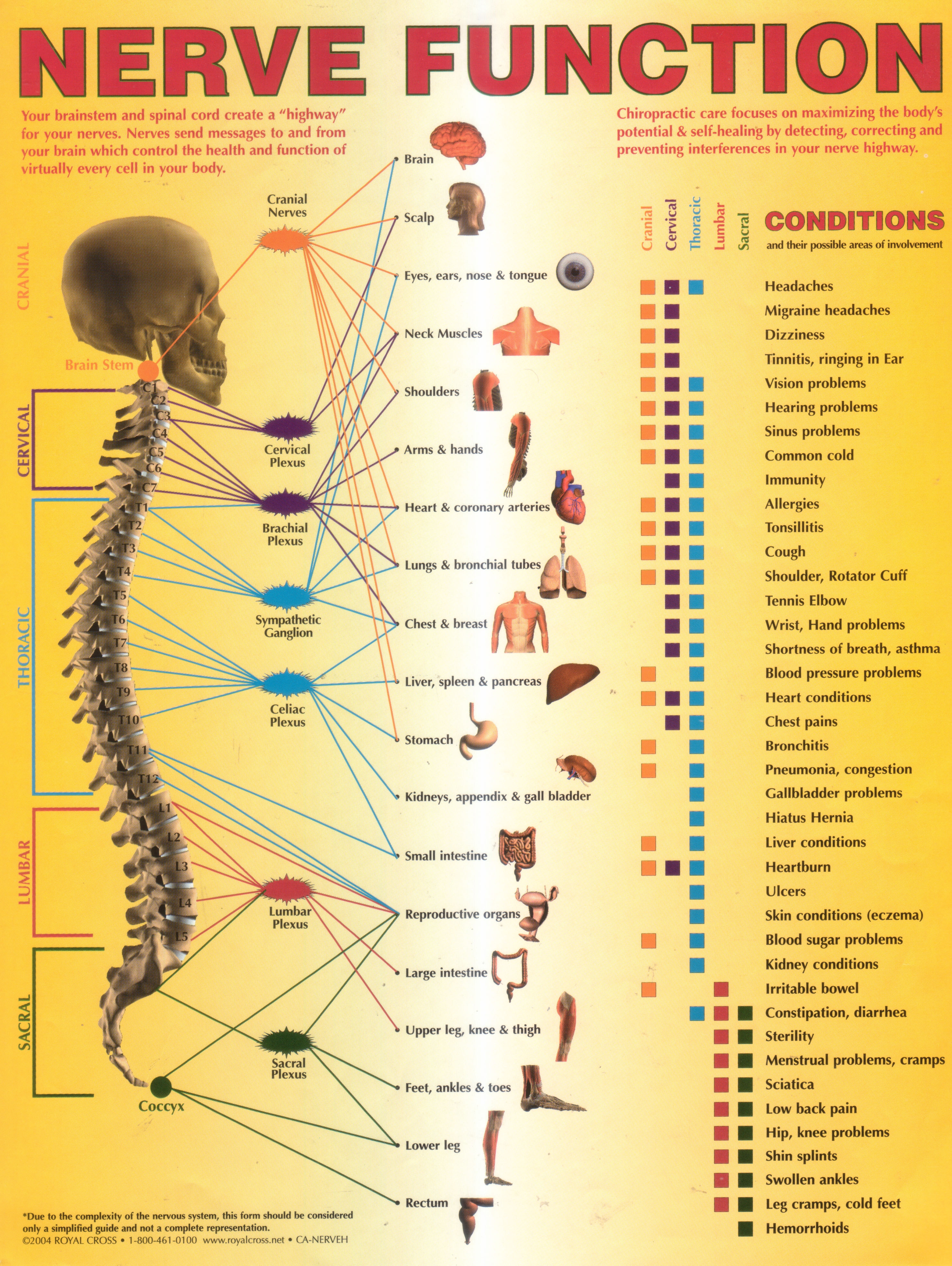
Annual World Spine Day Campaign Nerve Function Chart

Spinal Nerves Anatomical Chart Spine and Cranial Nervous System
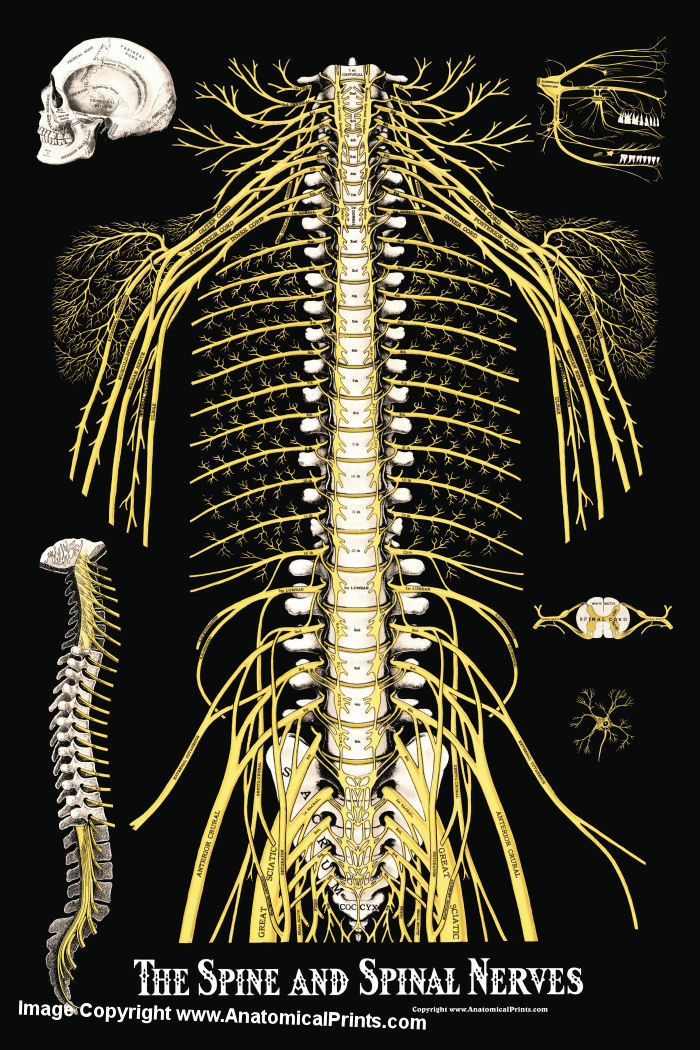
The Spine and Spinal Nerves Poster Clinical Charts and Supplies

Printable Spinal Nerve Chart

Lumbar Spinal Nerve Chart
These Nerves Exit The Intervertebral Foramina Below The Corresponding Vertebra.
The Peripheral Nerves Are Responsible For Sensations And Muscle Movements.
Web The Spinal Cord And Peripheral Nerves.
Each Of These Nerves Branches Out From The Spinal Cord, Dividing And Subdividing To Form A Network Connecting The Spinal Cord To Every Part Of The Body.
Related Post: