Spinal Nerve Innervation Chart
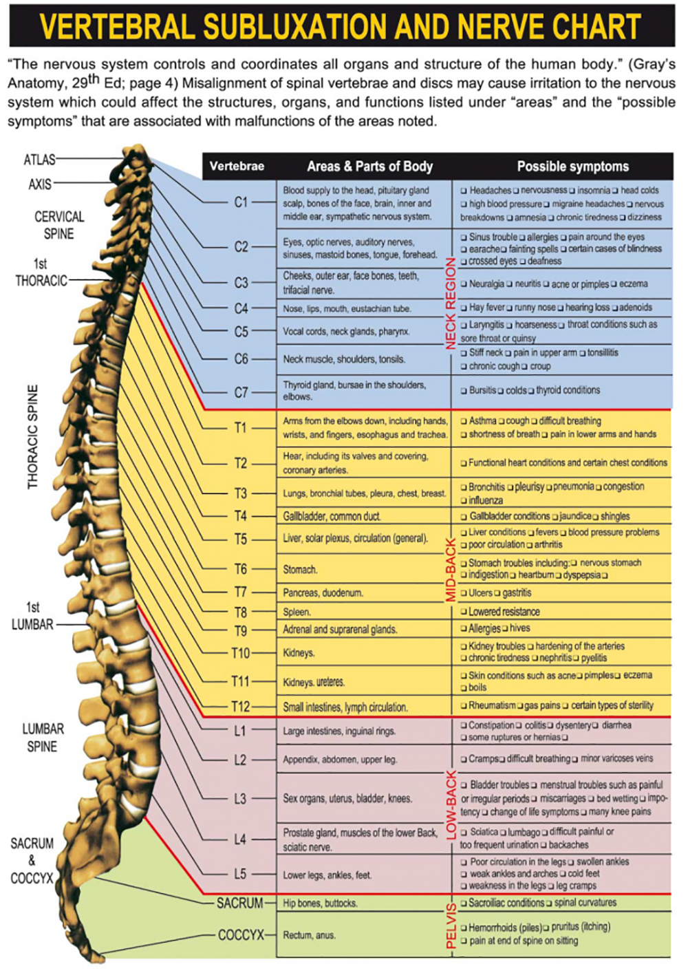
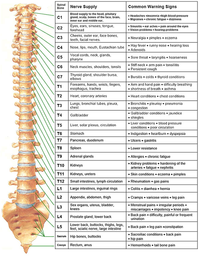
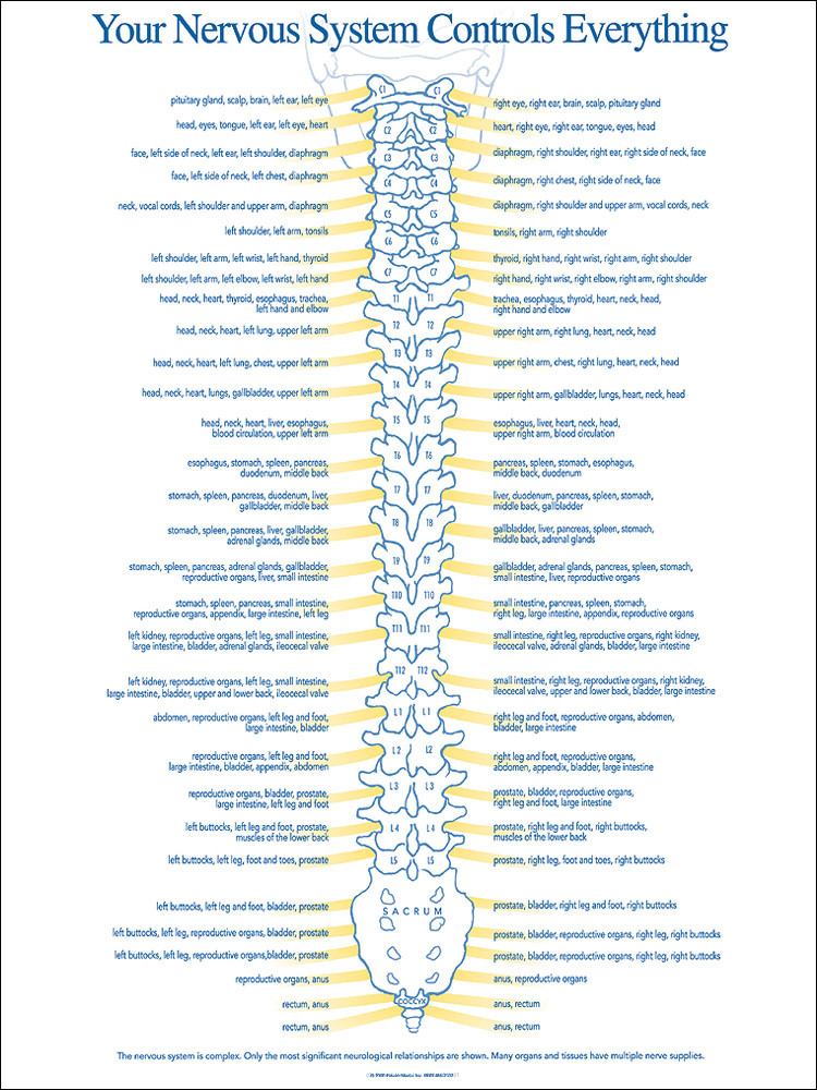
Spinal Nerve Innervation Chart - The chart depicts the intervertebral foramina between each vertebra where the spinal nerves exit. Outside the vertebral column, the nerve divides into branches. The roots connect via interneurons. Grossly, the root fibers join together within the intervertebral foramina to form a spinal nerve. Lumbar spinal nerves carry sensory and motor information to the lower body. Describe the sensory and motor components of spinal nerves and the plexuses that they pass through. A dermatome is an area of skin supplied by a single spinal nerve. Carries sensory (afferent) information to the spinal cord. Web the relevant anatomy of the innervation of the musculature of the back by the spinal nerves is centered around the lumbar spinal nerves, peripheral nerves of the lumbar plexus, spinal cord, and lumbar vertebral column. Web the 30 dermatomes explained and located. Web the relevant anatomy of the innervation of the musculature of the back by the spinal nerves is centered around the lumbar spinal nerves, peripheral nerves of the lumbar plexus, spinal cord, and lumbar vertebral column. Web these complex networks of nerves enable the brain to receive sensory inputs from the skin and to send motor controls for muscle movements.. A dermatome is an area of skin supplied by a single spinal nerve. Web by the end of this section, you will be able to: Numbers indicate the types of nerve fibers: For the most part, the spinal nerves exit the vertebral canal through the intervertebral foramen below their corresponding vertebra. This diagram indicates the formation of a typical spinal. Web the relevant anatomy of the innervation of the musculature of the back by the spinal nerves is centered around the lumbar spinal nerves, peripheral nerves of the lumbar plexus, spinal cord, and lumbar vertebral column. In general, the spinal cord consists of gray and white matter. It illustrates the cervical, thoracic, lumbar, and sacral regions of the spine and. The chart depicts the intervertebral foramina between each vertebra where the spinal nerves exit. In the cervical spine, there are eight pairs of spinal nerves labeled c1 to c8, which innervate the. This diagram indicates the formation of a typical spinal nerve from the dorsal and ventral roots. Eight cervical spinal nerve pairs, 12 thoracic pairs , five lumbar pairs,. Web the spinal nerve chart pdf is an anatomical diagram showing the nerves that exit from the spinal cord. Die spinalnerven sind ein wichtiger bestandteil des peripheren nervensystems (pns). Web many nerves come from the spinal cord, pass through foramina (holes) formed by notches of 24 vertebrae in the movable spinal column, and innervate or supply specific areas and parts. Web many nerves come from the spinal cord, pass through foramina (holes) formed by notches of 24 vertebrae in the movable spinal column, and innervate or supply specific areas and parts of the body.2 whenever specific areas or parts of the body are malfunctioning, generalized and/or specific symptoms are possible.3. Lumbar spinal nerves carry sensory and motor information to the. Describe the sensory and motor components of spinal nerves and the plexuses that they pass through. This diagram indicates the formation of a typical spinal nerve from the dorsal and ventral roots. The dorsal ramus contains nerves that serve the dorsal portions of the trunk; Each spinal nerve originates from two roots: In the cervical spine, there are eight pairs. The roots connect via interneurons. The chart depicts the intervertebral foramina between each vertebra where the spinal nerves exit. Web the spinal nerve chart pdf is an anatomical diagram showing the nerves that exit from the spinal cord. Spinal nerves can be impacted by a variety of medical conditions, resulting in pain, weakness, or decreased sensation. Grossly, the root fibers. Each spinal nerve originates from two roots: At each vertebral level, paired spinal nerves leave the spinal cord via the intervertebral foramina of the vertebral column. Web the spinal nerve chart pdf is an anatomical diagram showing the nerves that exit from the spinal cord. Web 12 nerve roots (t1 to t12) on each side of the spine that branch. Web each nerve forms from nerve fibers, known as fila radicularia, extending from the posterior (dorsal) and anterior (ventral) roots of the spinal cord. A dermatome is an area of skin supplied by a single spinal nerve. Each thoracic spinal nerve is named for the vertebra above it. Web many nerves come from the spinal cord, pass through foramina (holes). 8 cervical, 12 thoracic, 5 lumbar, 5 sacral, and 1 coccygeal, named according to their corresponding vertebral levels. Web muscle innervation reference tables. A dermatome is an area of skin supplied by a single spinal nerve. Eg the t4 nerve root runs between the t4 vertebra and t5 vertebra. A dermatome is a specific area of skin that is supplied by. In the cervical spine, there are eight pairs of spinal nerves labeled c1 to c8, which innervate the. Each of these nerves branches out from the spinal cord, dividing and subdividing to form a network connecting the spinal cord to every part of the body. Web there are 31 bilateral pairs of spinal nerves, named from the vertebra they correspond to. Die spinalnerven sind ein wichtiger bestandteil des peripheren nervensystems (pns). Eight cervical spinal nerve pairs, 12 thoracic pairs , five lumbar pairs, five sacral pairs, and one coccygeal. Grossly, the root fibers join together within the intervertebral foramina to form a spinal nerve. Web these complex networks of nerves enable the brain to receive sensory inputs from the skin and to send motor controls for muscle movements. The nerves connected to the spinal cord are the spinal nerves. Web each nerve forms from nerve fibers, known as fila radicularia, extending from the posterior (dorsal) and anterior (ventral) roots of the spinal cord. Web 12 nerve roots (t1 to t12) on each side of the spine that branch from the spinal cord. Each spinal nerve originates from two roots:
Spinal Nerve Function Anatomical Chart Anatomy Models and Anatomical

Printable Spinal Nerve Chart Free Printable Calendar

Spinal Nerve Chart medschool doctor medicalstudent Image Credits

Blog Best Rated Chiropractor in New York

Cervical Nerves Innervation

Lumbar Spinal Nerve Chart

CONDITIONS Sault Chiropractors

Pin on Health & Wellness Tips

Chiropractic Spinal Nerve Chart Nerve Function Chart

spinal nerves innervation
Web How To Use The Spinal Nerve Chart:
In General, The Spinal Cord Consists Of Gray And White Matter.
Each Nerve Then Divides Into Anterior And Posterior Nerve Fibres.
Outside The Vertebral Column, The Nerve Divides Into Branches.
Related Post: