Teeth Anatomy Chart
Teeth Anatomy Chart - The pulp, dentin, and enamel. Premolars are only present in the permanent dentition. Web dental charts are normally arranged from the viewpoint of a dental practitioner facing a patient. Prefer to learn by doing? Web learn about the types of teeth in a fast and efficient way using our interactive tooth identification quizzes and labeled diagrams. Look no further than our dental anatomy quizzes and tooth diagrams. Dental anatomy is a field of anatomy dedicated to the study of tooth structure. Your teeth play a big role in digestion. These teeth are shaped like chisels and are good at biting off small bits of food. Fully labeled illustrations of the teeth with dental terminology (orientation, surfaces, cusps, roots numbering systems) and detailed images of each permanent tooth. The upper teeth are often referred to as the maxillary teeth, and are found along the alveolar ridge of the maxilla. Fully labeled illustrations of the teeth with dental terminology (orientation, surfaces, cusps, roots numbering systems) and detailed images of each permanent tooth. There are 8 incisors in both the permanent and primary dentition, with four in each dental arch.. Web a teeth chart is a simple drawing or illustration of your teeth with names, numbers, and types of teeth. The development, appearance, and classification of teeth fall within its field of study, though dental occlusion, or contact between teeth, does not. The upper teeth are often referred to as the maxillary teeth, and are found along the alveolar ridge. Your teeth play a big role in digestion. These teeth are referred to as letters a, b, c, d and e. Web learn about the types of teeth in a fast and efficient way using our interactive tooth identification quizzes and labeled diagrams. The pulp, dentin, and enamel. Web starting at the front of the mouth, in the center, there. Fully labeled illustrations of the teeth with dental terminology (orientation, surfaces, cusps, roots numbering systems) and detailed images of each permanent tooth. Web this dental anatomy chart provides a comprehensive and lifelike representation of permanent human teeth, including incisors, canines, premolars, and molars. Enamel, which is the hardest substance in the body, is on the outside of the tooth. Use. The development, appearance, and classification of teeth fall within its field of study, though dental occlusion, or contact between teeth, does not. The patient's right side appears on the left side of the chart, and the patient's left side appears on the right side of the chart. Web each tooth is an organ consisting of three layers: Web learn about. The development, appearance, and classification of teeth fall within its field of study, though dental occlusion, or contact between teeth, does not. The names are given to these surfaces according to their position and use. Web starting at the front of the mouth, in the center, there are the central incisors and then the lateral incisors. It serves as a. Premolars are only present in the permanent dentition. Web a teeth chart is a simple drawing or illustration of your teeth with names, numbers, and types of teeth. The permanent dentition is composed of 32 teeth with 16 in each arch. The anterior teeth consist of the incisors and canine teeth, and the posterior teeth consist of the premolars and. There are four main types of teeth in humans, shown labelled here. The pulp of the tooth is a vascular region of soft connective tissues in the middle of the tooth. The first thing to know is that most normal adult mouths contain 32 teeth. What are teeth made of? Web atlas of dental anatomy: Web learn about the types of teeth in a fast and efficient way using our interactive tooth identification quizzes and labeled diagrams. Web the teeth are categorized as incisors, canines, premolars, and molars and conventionally are numbered beginning with the maxillary right third molar (see figure identifying the teeth). The crown of the tooth is what is visible in the. Web the teeth are categorized as incisors, canines, premolars, and molars and conventionally are numbered beginning with the maxillary right third molar (see figure identifying the teeth). Most people start off adulthood with 32 teeth, not including the wisdom teeth. Fully labeled illustrations of the teeth with dental terminology (orientation, surfaces, cusps, roots numbering systems) and detailed images of each. Web starting at the front of the mouth, in the center, there are the central incisors and then the lateral incisors. Primary (baby) teeth are usually replaced by adult teeth between the ages of 6 and 12. Web the anatomy of a tooth divides into two main sections: The labels right and left on the charts in this article correspond to the patient's right and left, respectively. Look no further than our dental anatomy quizzes and tooth diagrams. There are separate teeth number charts for adults as well as babies. The names are given to these surfaces according to their position and use. Web teeth diagrams labeled and unlabeled. Tiny blood vessels and nerve fibers enter the pulp through small holes in the tip of the roots to support the hard outer structures. This diagram helps us learn the names of each tooth, the. The primary teeth begin to erupt at 6 months of age. There are four main types of teeth in humans, shown labelled here. Web (top) growing of tooth. Web the five surfaces are labial, palatal, mesial, distal and incisal surfaces. The second layer is dentin, which. The numbering system shown is the one most commonly used in the united states.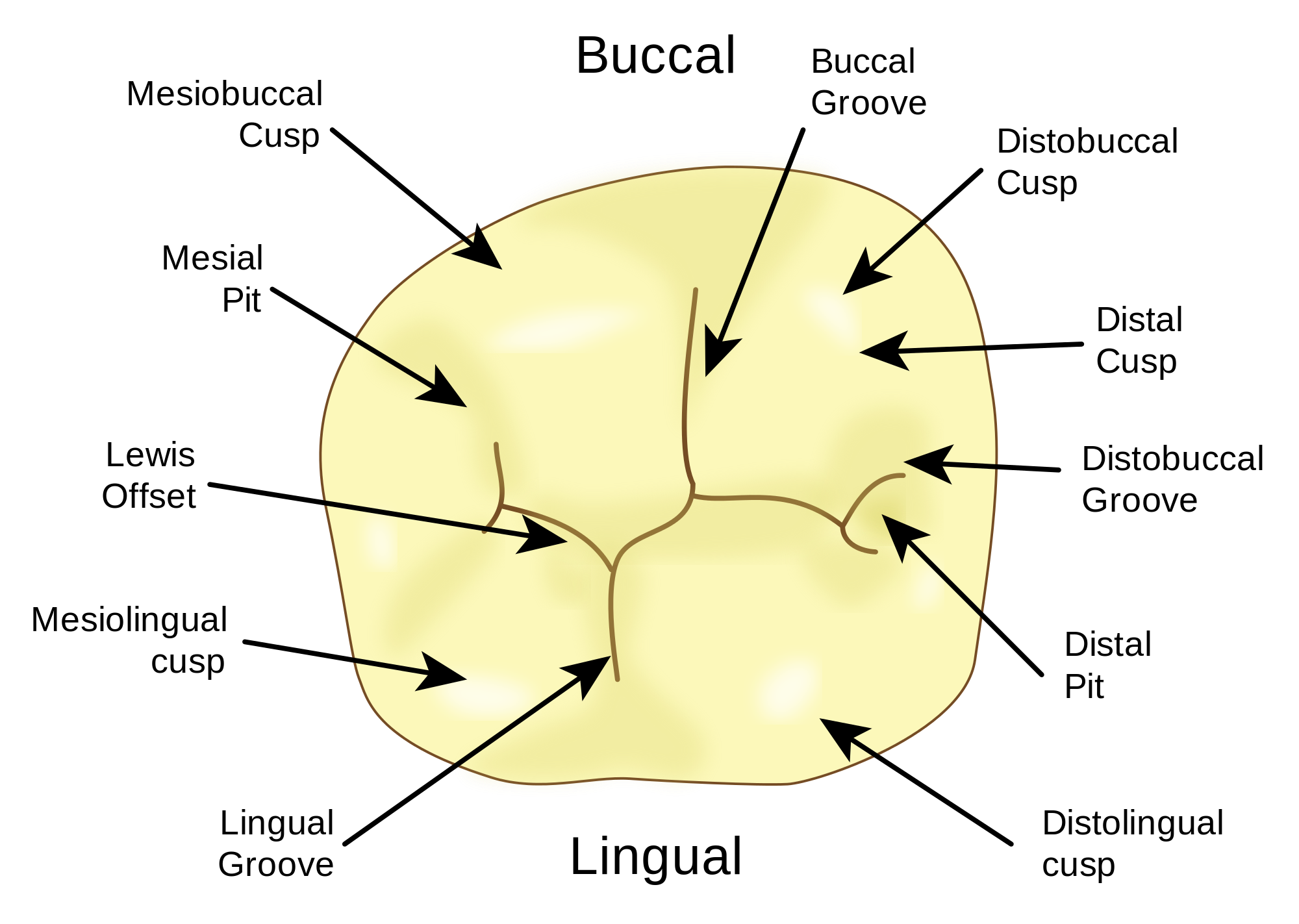
Your Tooth Surfaces Explained Dental Clinique
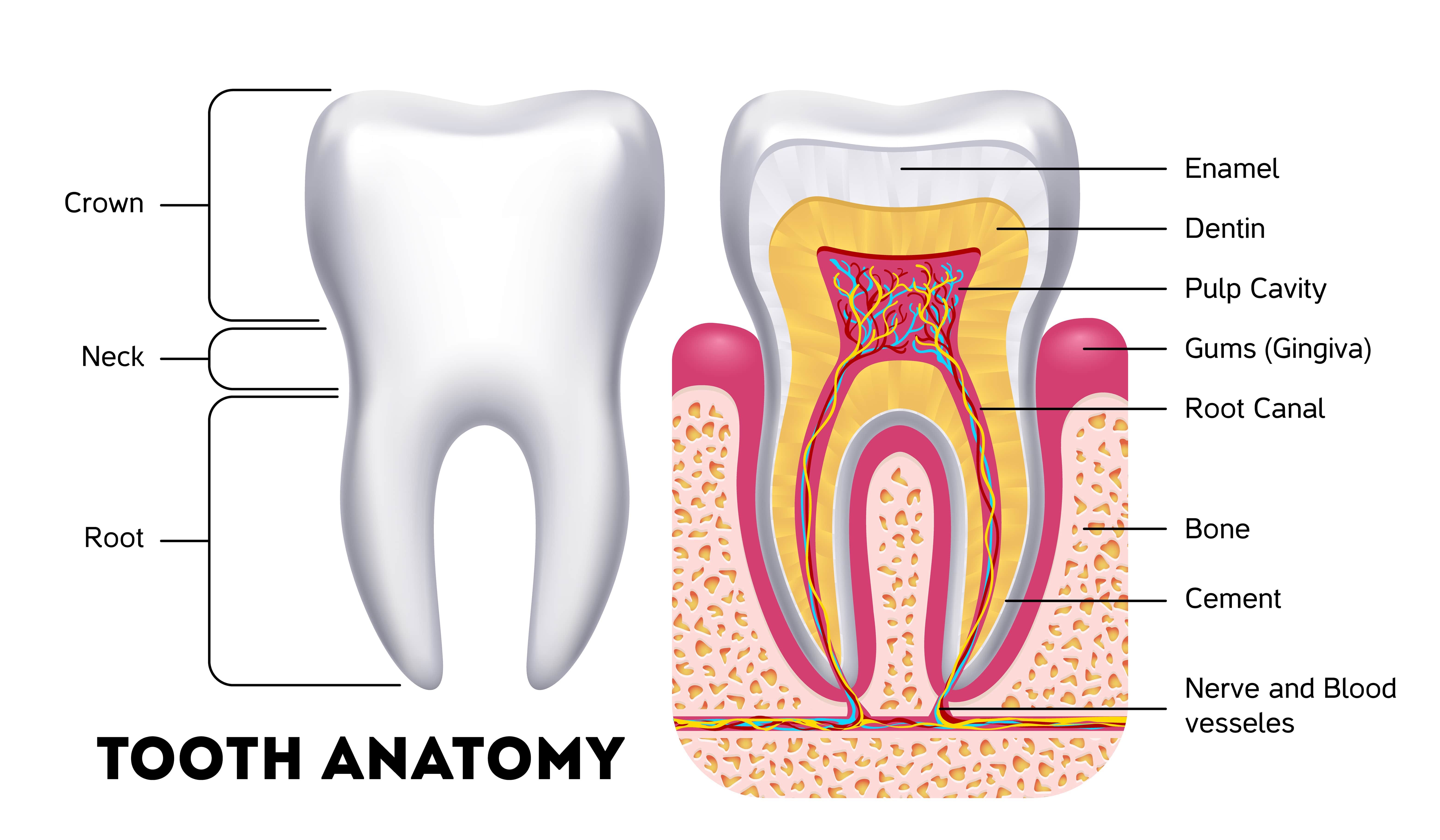
Anatomy Of The Teeth Anatomical Chart Poster Prints Images and Photos
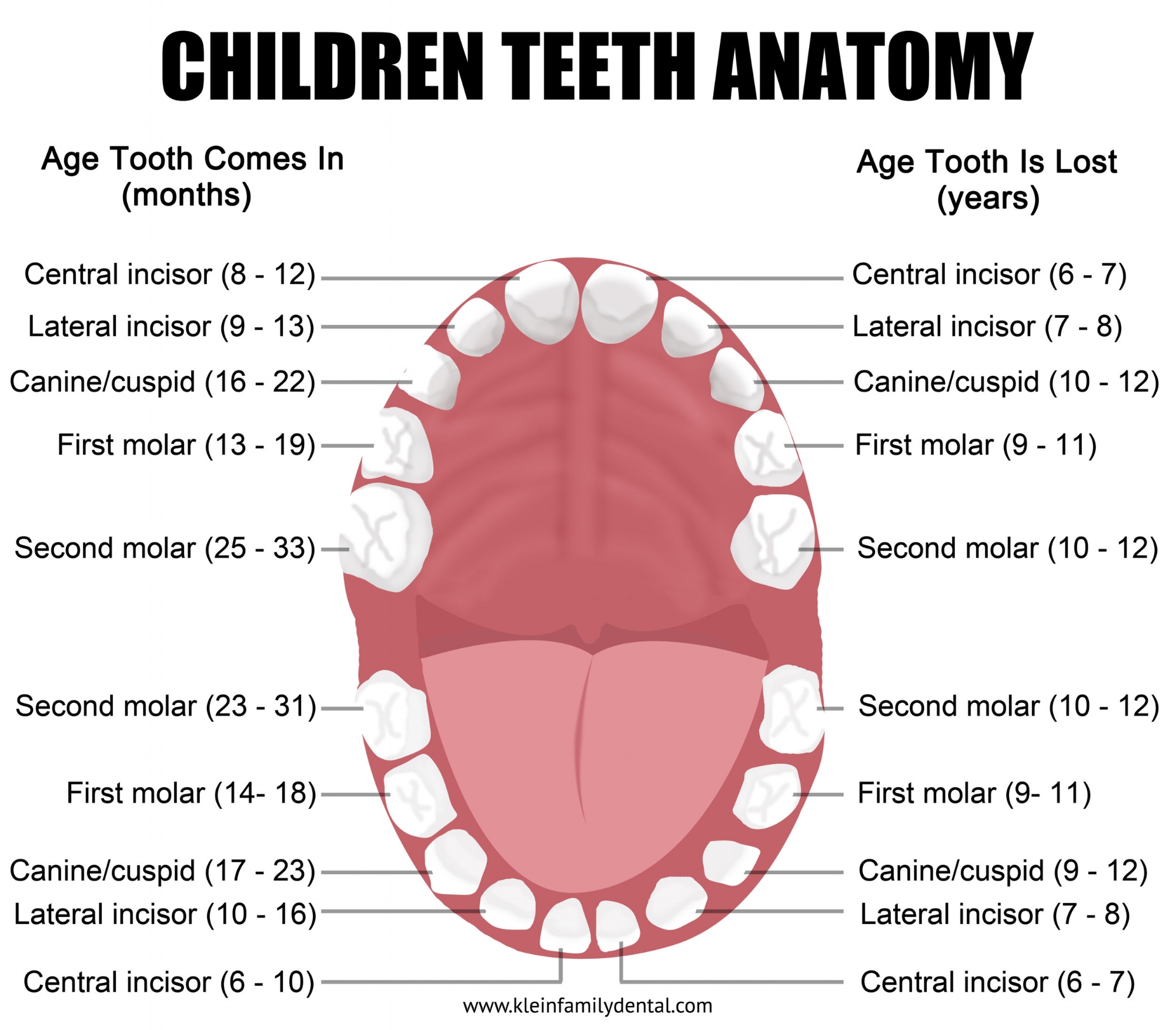
Pediatric Tooth Chart — Klein Family Dental
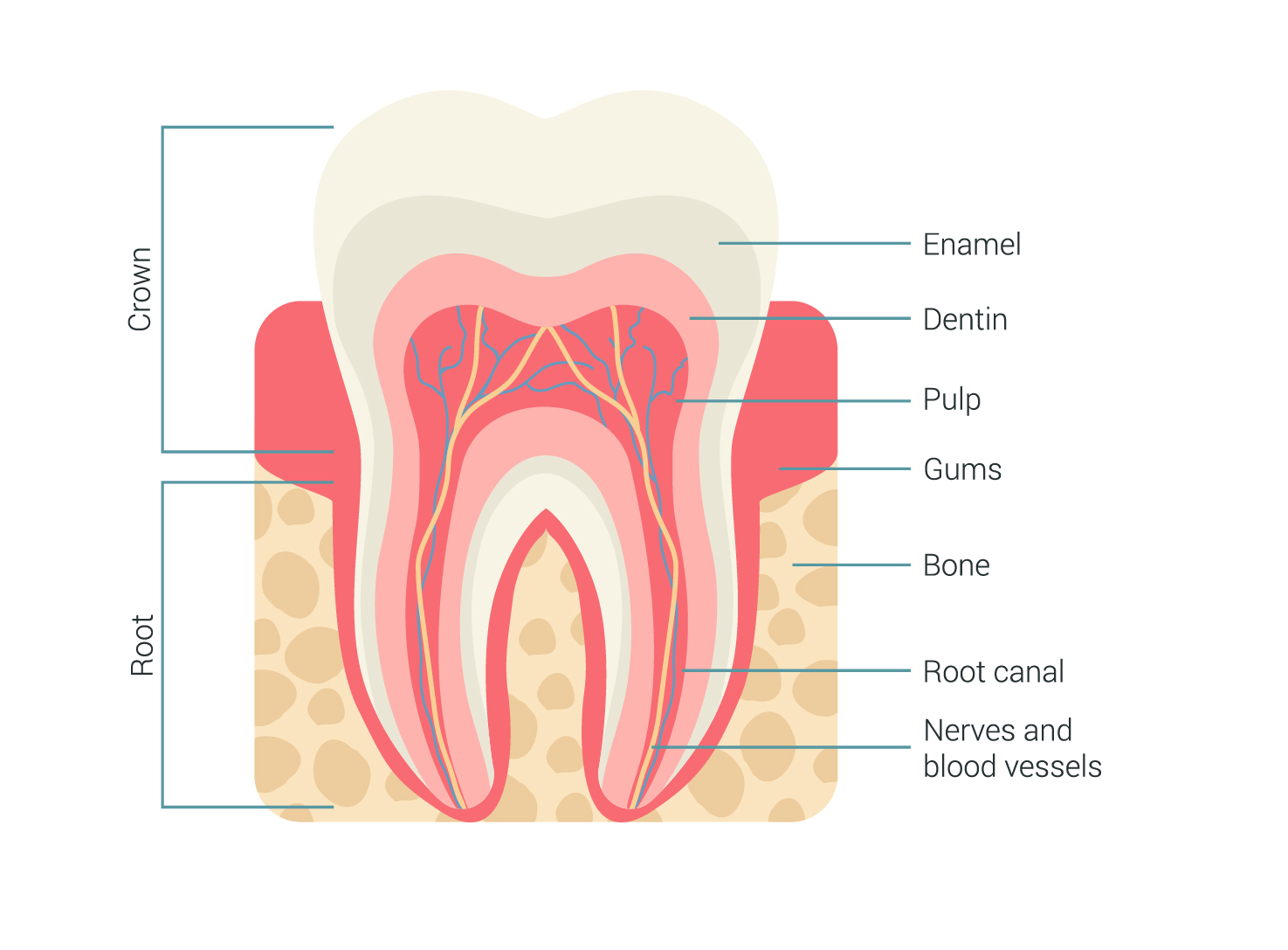
Tooth Anatomy Milford Family Dentistry

Anatomy of The Teeth Anatomical Chart 20'' x 26''
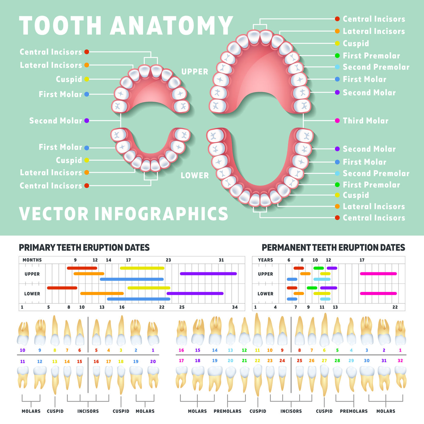
Orthodontist human tooth anatomy vector infographics with teeth diagra

Child and Adult Dentition (Teeth) Structure Primary Permanent

Tooth Numbers and illustrations

The Different Types of Teeth Gentle Dentist

Teeth Anatomy Chart Australia
Web Teeth Are Made Up Of Different Layers — Enamel, Dentin, Pulp, And Cementum.
Dental Anatomy Is A Field Of Anatomy Dedicated To The Study Of Human Tooth Structures.
The Development, Appearance, And Classification Of.
Read On To Find Out.
Related Post: