X Ray Exposure Chart
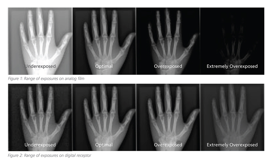
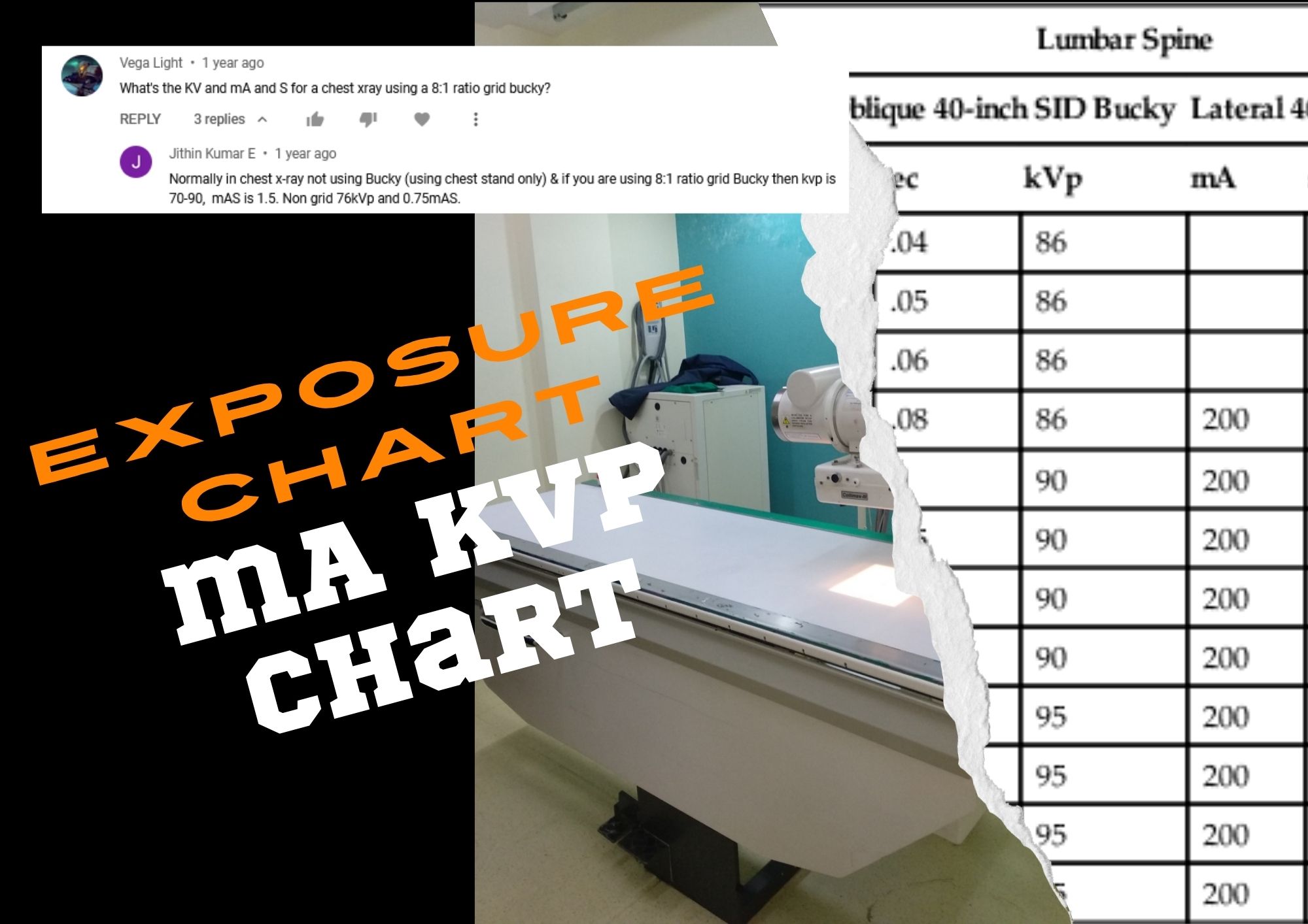
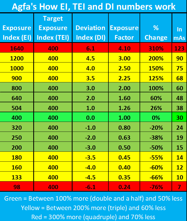
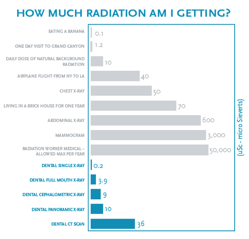
X Ray Exposure Chart - Patients want to know if radiation from mammograms, bone density tests, computed tomography (ct) scans, and so forth will increase their risk of developing cancer. The body part imaged (hand, foot, skull, etc) the kv or kilovolts required for the image (how strong of a beam) Web with the free download: • explain the relationship between milliamperage and exposure time with radiation production and image receptor exposure. Grid mas cm kvp mas cm kvp mas cm kvp. Waters y 40 80 44. The exposure chart is applied for: Web acr recommendations and resources designed to assist radiologists in providing effective imaging and therapy while minimizing the risk during exposure to ionizing radiation. Web the technical parameters which control the exposure measured on the detector and also the radiation dose delivered to the patient are summarized in the table below and include (ma, kvp, s, sid and bucky factor). This chart simplifies a highly complex topic for patients’ informational use. There's always questions about radiation exposure from medical imaging. Web ct, radiography, and fluoroscopy all work on the same basic principle: This chart simplifies a highly complex topic for patients’ informational use. Effective dose allows your doctor to evaluate your risk and compare it to common, everyday sources of exposure, such as natural background radiation. Web acr recommendations and resources. Web acr recommendations and resources designed to assist radiologists in providing effective imaging and therapy while minimizing the risk during exposure to ionizing radiation. The uv index provides a daily forecast of the expected intensity of uv radiation from the sun. Grid mas cm kvp mas cm kvp mas cm kvp. • calculate changes in milliamperage and exposure time to. E = q / m where e is exposure, q is the quantity of charge on the ions and m is the unit mass of air. The production of an exposure chart calls for either a large step wedge or a series of plates of different thicknesses made from the same material to which the chart relates. Waters y 40. Web radiographic exposure technique. Web these are average exposures using a carestream cr system, exposures may vary dramatically between different film/screen combinations, cr or dr systems. Many diagnostic exposures are less than or similar to the exposure we receive from natural background radiation. The exposure chart is applied for: Waters y 40 80 44. The uv index provides a daily forecast of the expected intensity of uv radiation from the sun. Install the uv index app on your mobile device: Web a standard radiography technique chart is a written table that contains the following technical data to help radiographers obtain a consistent, standardized image while using the lowest radiation dose possible: • explain the. A given density, for example: Effective dose allows your doctor to evaluate your risk and compare it to common, everyday sources of exposure, such as natural background radiation. Web acr recommendations and resources designed to assist radiologists in providing effective imaging and therapy while minimizing the risk during exposure to ionizing radiation. Waters y 40 80 44. The actual dose. This document summarizes the development of an exposure chart to optimize digital radiography techniques for pediatric patients at the royal children's hospital in brisbane, australia. A millisievert is a measure of radiation dose which accounts. Waters y 40 80 44. Small medium large small medium large. Web ct, radiography, and fluoroscopy all work on the same basic principle: The uv index provides a daily forecast of the expected intensity of uv radiation from the sun. This document summarizes the development of an exposure chart to optimize digital radiography techniques for pediatric patients at the royal children's hospital in brisbane, australia. Effective dose allows your doctor to evaluate your risk and compare it to common, everyday sources of exposure,. The actual dose can vary substantially, depending on a person’s size as well as on differences in imaging practices. Small medium large small medium large. • explain the relationship between milliamperage and exposure time with radiation production and image receptor exposure. Grid mas cm kvp mas cm kvp mas cm kvp. Web in the uk, public health england calculated that. Web the uv index. Grid mas cm kvp mas cm kvp mas cm kvp. The uv index provides a daily forecast of the expected intensity of uv radiation from the sun. Web the technical parameters which control the exposure measured on the detector and also the radiation dose delivered to the patient are summarized in the table below and include. Learn more about how the uv index can be calculated and analyzed and sign up for uv index email alerts. A given density, for example: Web a standard radiography technique chart is a written table that contains the following technical data to help radiographers obtain a consistent, standardized image while using the lowest radiation dose possible: • explain the relationship between milliamperage and exposure time with radiation production and image receptor exposure. The aim of this review was to develop a radiographic optimisation strategy to make use of digital. The production of an exposure chart calls for either a large step wedge or a series of plates of different thicknesses made from the same material to which the chart relates. Web with the free download: Many diagnostic exposures are less than or similar to the exposure we receive from natural background radiation. The exposure chart is applied for: Join our webinar and gain insights from industry experts at imv imaging. Web the technical parameters which control the exposure measured on the detector and also the radiation dose delivered to the patient are summarized in the table below and include (ma, kvp, s, sid and bucky factor). A millisievert is a measure of radiation dose which accounts. Small medium large small medium large. This document summarizes the development of an exposure chart to optimize digital radiography techniques for pediatric patients at the royal children's hospital in brisbane, australia. Web ct, radiography, and fluoroscopy all work on the same basic principle: The uv index provides a daily forecast of the expected intensity of uv radiation from the sun.
A paediatric Xray exposure chart Semantic Scholar

Understanding Radiology Exposure Indicators Everything Rad

What To Know About Dental XRays

A paediatric Xray exposure chart Semantic Scholar

Dental Radiation Exposure Comparison Chart

Relevance of Exposure Chart with HighFrequency XRay Machine

Exposure parameters and radiographic technique for selected Xray

What Is Exposure Index In Digital Radiography

Radiographic Exposure Chart

Xray Exposure Chart
This Chart Simplifies A Highly Complex Topic For Patients’ Informational Use.
The Body Part Imaged (Hand, Foot, Skull, Etc) The Kv Or Kilovolts Required For The Image (How Strong Of A Beam)
The Actual Dose Can Vary Substantially, Depending On A Person’s Size As Well As On Differences In Imaging Practices.
Web These Are Average Exposures Using A Carestream Cr System, Exposures May Vary Dramatically Between Different Film/Screen Combinations, Cr Or Dr Systems.
Related Post: