X Ray Positioning Chart
X Ray Positioning Chart - Web detailed instructions for more than 200 radiographic positions: Image receptor size and orientation. You can use it to image animals in a veterinary setting and humans in a medical setting. Web radiographic positioning terminology is used routinely to describe the position of the patient for taking various radiographs. Visualize how the image would look on a monitor. So, what is a technique. Purpose and structures shown clear image of entire pelvis. Anterior denotes the front of a body part, while the posterior denotes the back. Web what is the purpose of a radiography techniques chart? The lungs and spine should be visible behind the heart shadow. Superior denotes the top of a body part, while inferior denotes the bottom. For each setup in the tables, there is a picture. The patient is supine (on an inclined radiographic table) with the head lower than the feet. The base of the skull is complex and consists of the paired temporal bones as well as the sphenoid and ethmoid.. Purpose and structures shown clear image of entire pelvis. Move the patient and position the area of interest along the long axis of your collimated field, rather than rotating the collimator. The base of the skull is complex and consists of the paired temporal bones as well as the sphenoid and ethmoid. The front of the cranium consists of the. Anterior denotes the front of a body part, while the posterior denotes the back. Move the patient and position the area of interest along the long axis of your collimated field, rather than rotating the collimator. Web what is the purpose of a radiography techniques chart? The front of the cranium consists of the frontal bone. • visualize how the. Web detailed instructions for more than 200 radiographic positions: Proper positioning ensures that the imaging equipment captures clear and accurate images of the targeted area, while also minimizing radiation exposure to the patient and healthcare professionals. Positioning decision support function ai *2 supports general radiography operations. Image receptor size and orientation. Visualize how the image would look on a monitor. Positioning decision support function ai *2 supports general radiography operations. The base of the skull is complex and consists of the paired temporal bones as well as the sphenoid and ethmoid. This chart is vital in the medical imaging field. Position of patient supine position. • visualize how the image would look on a monitor. Move the patient and position the area of interest along the long axis of your collimated field, rather than rotating the collimator. Correct positioning isn’t just crucial for diagnostic accuracy and minimizes the patient’s exposure to radiation. • visualize how the image would look on a monitor. There are myriad ways to enhance your skills, from websites and apps to. Web understanding patient positioning requires a knowledge of the basic terminology relating to radiographic positioning: Both costophrenic angles and the lower parts of the diaphragm should be visible. Web what is the purpose of a radiography techniques chart? Superior denotes the top of a body part, while inferior denotes the bottom. Purpose and structures shown clear image of entire pelvis. Suggested digital imaging systems technical factor settings for all positions (except nephrotomography) more about techniques. Positioning decision support function ai *2 supports general radiography operations. Anterior denotes the front of a body part, while the posterior denotes the back. Standard nomenclature is employed with respect to the anatomic position. The lungs and spine should be visible behind the heart shadow. Anterior denotes the front of a body part, while the posterior denotes the back. Web the exposure should be made at full inspiration and should show rib 10 posteriorly above the diaphragm and rib 6 anteriorly. Superior denotes the top of a body part, while inferior denotes the bottom. There are myriad ways to enhance your skills, from websites and. Suggested digital imaging systems technical factor settings for all positions (except nephrotomography) more about techniques. The patient is supine (on an inclined radiographic table) with the head lower than the feet. Web radiographic positioning terminology is used routinely to describe the position of the patient for taking various radiographs. There are myriad ways to enhance your skills, from websites and. Image receptor size and orientation. The base of the skull is complex and consists of the paired temporal bones as well as the sphenoid and ethmoid. Visualize how the image would look on a monitor. Move the patient and position the area of interest along the long axis of your collimated field, rather than rotating the collimator. Proper positioning ensures that the imaging equipment captures clear and accurate images of the targeted area, while also minimizing radiation exposure to the patient and healthcare professionals. Suggested digital imaging systems technical factor settings for all positions (except nephrotomography) more about techniques. Web the right and left parietal bones and right and left temporal bones make up the sides of the cranium. It equips professionals with the knowledge needed to avoid common positioning mistakes, ensuring that the images obtained are of the highest quality for accurate diagnosis. Web detailed instructions for more than 200 radiographic positions: Right side touches the cassette. This chart is vital in the medical imaging field. For each setup in the tables, there is a picture. Web radiographic positioning terminology is used routinely to describe the position of the patient for taking various radiographs. Web understanding patient positioning requires a knowledge of the basic terminology relating to radiographic positioning: You can use it to image animals in a veterinary setting and humans in a medical setting. So, what is a technique.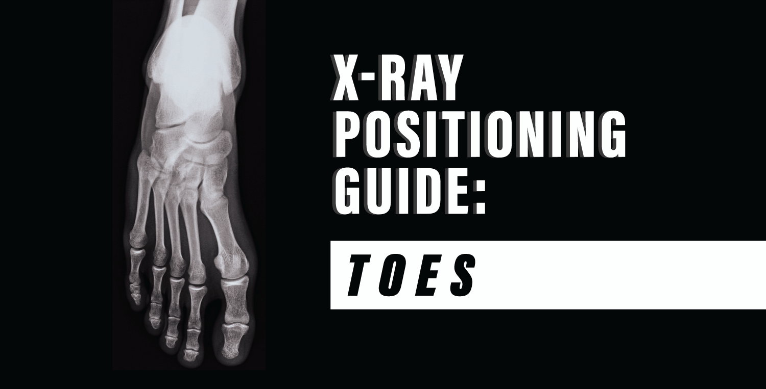
XRay Positioning Guide Toes Medical Professionals

Checking Invasive Devices Using Chest XRays Dr. GrepMed
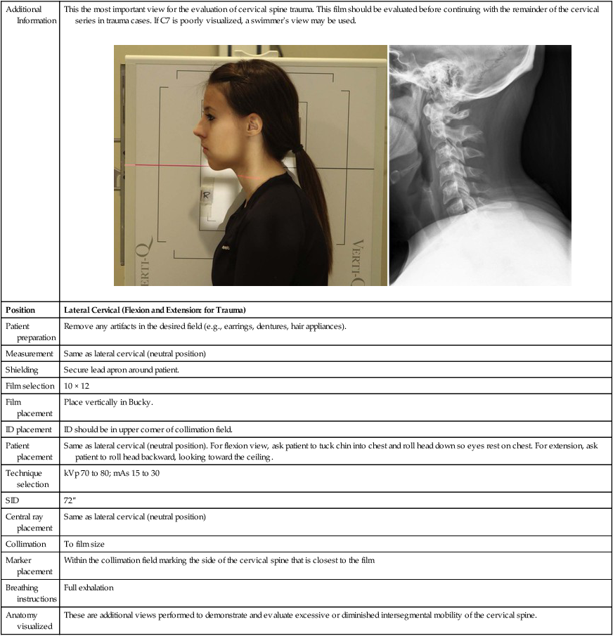
Radiographic Positioning Radiology Key
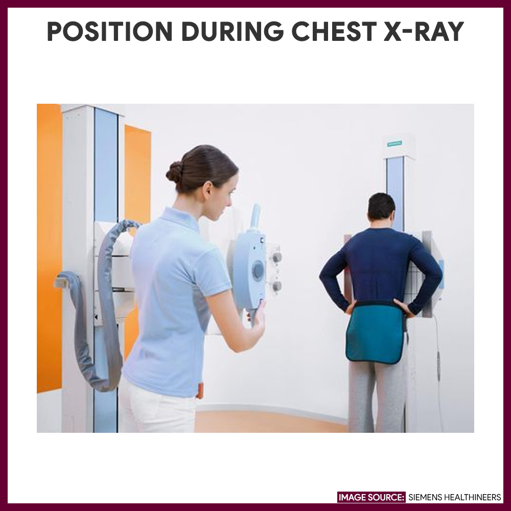
Chest Xray (Chest Radiography) Nursing Responsibilities Nurseslabs
Radiographic Positioning Chart PDF
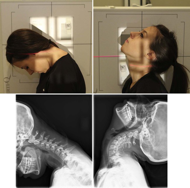
Radiographic Positioning Radiology Key
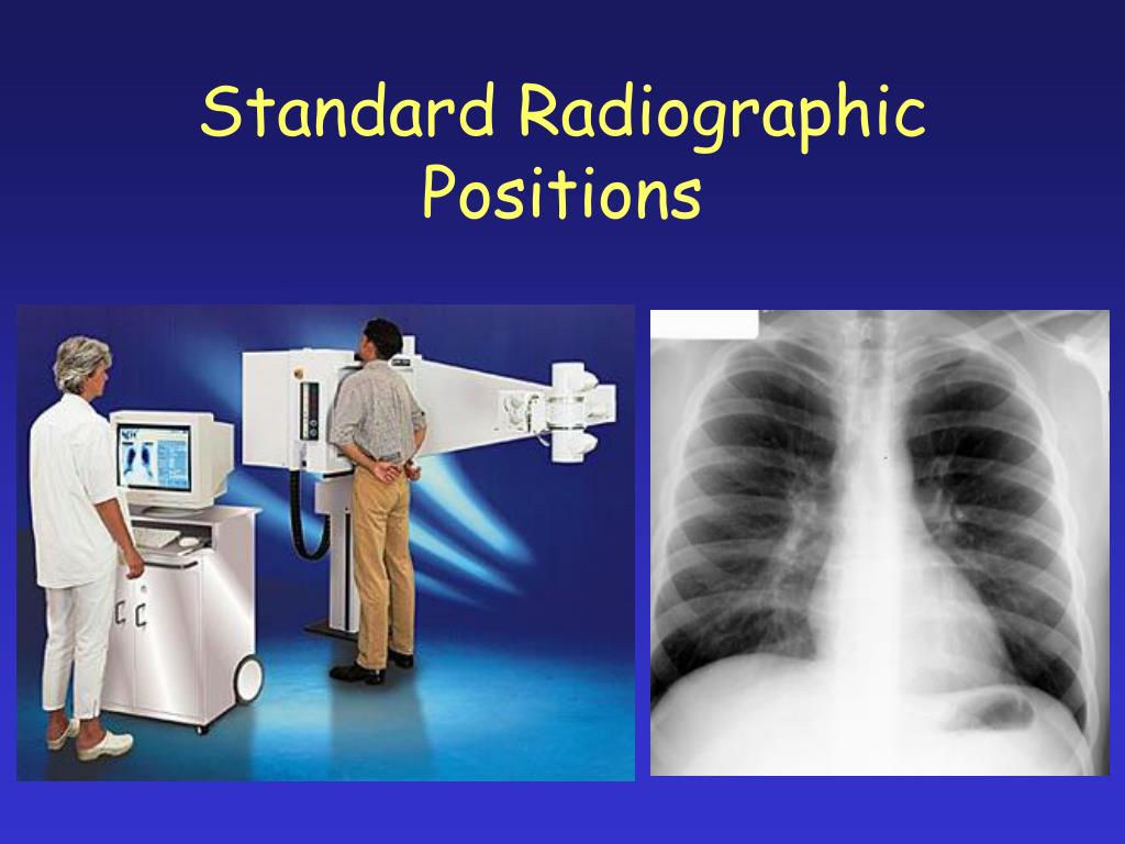
X Ray Position Chart

Skull X Ray Positioning Chart
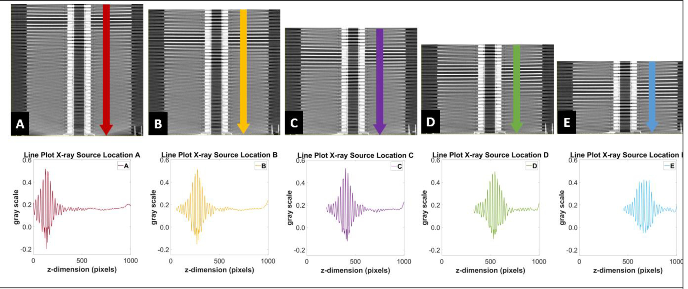
X Ray Positioning Chart Free Download evermagic
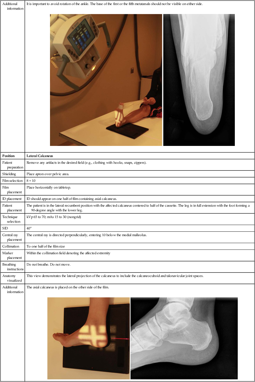
Radiographic Positioning Radiology Key
Standard Nomenclature Is Employed With Respect To The Anatomic Position.
Position Of Patient Supine Position.
Correct Positioning Isn’t Just Crucial For Diagnostic Accuracy And Minimizes The Patient’s Exposure To Radiation.
• Visualize How The Image Would Look On A Monitor.
Related Post:
