Frog Dissection Drawing
Frog Dissection Drawing - Place the frog in the dissecting pan ventral side up. Web obtain a preserved frog and place it on your dissecting tray, dorsal surface up. If your class is going to dissect a frog, your teacher should provide all the necessary tools for the job. Make transverse (horizontal) cuts near the arms and legs. Digestive system of the frog anatomy model. One of the best ways to learn about adult amphibian anatomy is to dissect a preserved frog and see how all the organs fit together inside its body. The frog has no neck so the trunk follows immediately behind the head. Why is the color and texture the way that it is? The fleshy upper eyelid, the thinner lower eyelid, and the transparent nictitating membrane. Along with toads, they make up the largest group of amphibians. What do you notice about it? Frogs and other small animals are typically dissected in biology labs to learn anatomy. On the outside of the frog’s head are two external nares, or nostrils; Along with toads, they make up the largest group of amphibians. As members of the class amphibia, frogs may live some of their adult lives on land,. Frogs are vertebrates in a class called amphibians. In this lab, as we continue our dissection, we will take a close look at the frog’s. Web opal art 5.89k subscribers subscribe 2.7k views 1 year ago 10th biology federal board fsc, biology practical copy, experiment 25 indentification of different parts of the respiratory and. It can also be a great. Frogs have similar body systems (like the digestive and circulatory systems) to other vertebrates like humans, making them a nice specimen for comparative anatomy. During the dissection, students typically examine the organs, including the heart, lungs, liver, stomach, and intestines. Web 1 prepare the dissection tray and get your frog. Web place the frog in the dissecting pan ventral side. Place the frog in the dissecting pan ventral side up. Web frog dissection is a process of examining the internal anatomy of a frog by making cuts through the skin and muscles. Life the flaps of the body wall and pin back. Web 1 prepare the dissection tray and get your frog. Cut along the midline of the body to. Web obtain a preserved frog and place it on your dissecting tray, dorsal surface up. Notice the appendages developed for a terrestrial life. If your class is going to dissect a frog, your teacher should provide all the necessary tools for the job. Use this guide for complete instructions. During the dissection, students typically examine the organs, including the heart,. Eggs are laid and fertilized in water. Web place the frog in the dissecting pan ventral side up. The frog has three eyelids: Place the frog in the dissecting pan ventral side up. Frogs and other small animals are typically dissected in biology labs to learn anatomy. Use scissors to lift the abdominal muscles away from the body cavity. As members of the class amphibia, frogs may live some of their adult lives on land, but they must return to water to reproduce. Cut along the midline of the body to the forelimbs. Draw the external anatomy of your frog. And two eyes, each of which has. During the dissection, students typically examine the organs, including the heart, lungs, liver, stomach, and intestines. Web if your classroom doesn’t have access to frog dissection specimens, visible biology ’s interactive 3d frog model can walk students through frog anatomy without prep or cleanup. Observe that each forelimb is divided into an upper arm, forearm, and hand. Draw the external. Web lesson plan for the frog dissection. Life the flaps of the body wall and pin back. The fleshy upper eyelid, the thinner lower eyelid, and the transparent nictitating membrane. Place the frog in the dissecting pan ventral side up. Web the app features four dissectible animal models: Includes a list of terms to study for lab practical. Make transverse (horizontal) cuts near the arms and legs. Digestive system of the frog anatomy model. Use scissors to lift the abdominal muscles away from the body cavity. Frogs are vertebrates in a class called amphibians. Frogs have similar body systems (like the digestive and circulatory systems) to other vertebrates like humans, making them a nice specimen for comparative anatomy. Draw the external anatomy of your frog. Mouth with opening to esophagus and trachea; Web the app features four dissectible animal models: The nictitating membrane is drawn up over the eye when the frog is underwater. Basic line drawings that students can practice labeling. If your class is going to dissect a frog, your teacher should provide all the necessary tools for the job. Frogs are vertebrates in a class called amphibians. Use scissors to lift the abdominal muscles away from the body cavity. Products used in this video:frog specimen: Frogs are vertebrates in a class called amphibians. Make transverse (horizontal) cuts near the arms and legs. Web frog dissection diagram and labeling go back to: Cut along the midline of the body to the forelimbs. On the outside of the frog’s head are two external nares, or nostrils; Use scissors to lift the abdominal muscles away from the body cavity.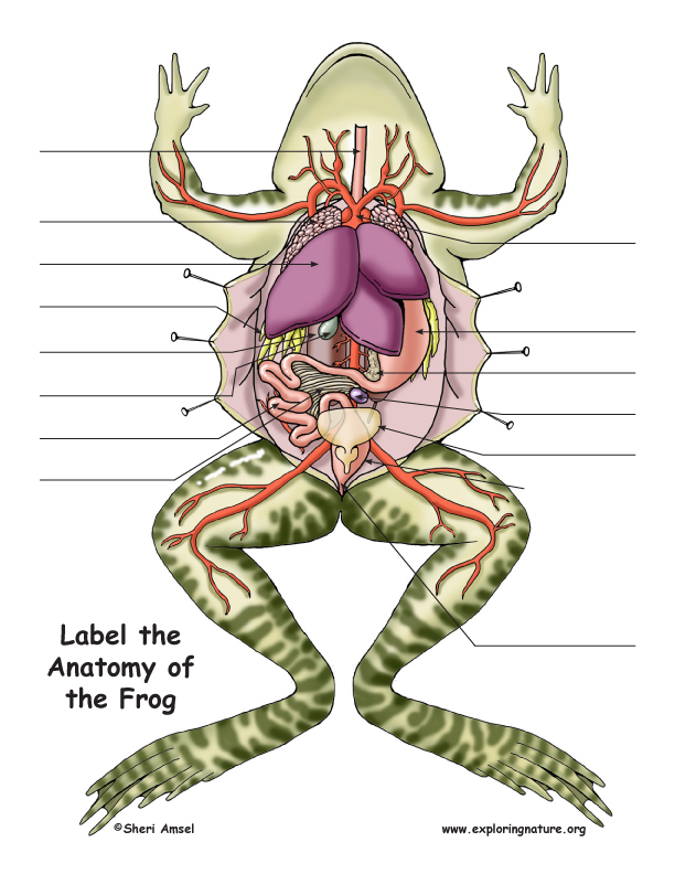
Frog Dissection Diagram and Labeling
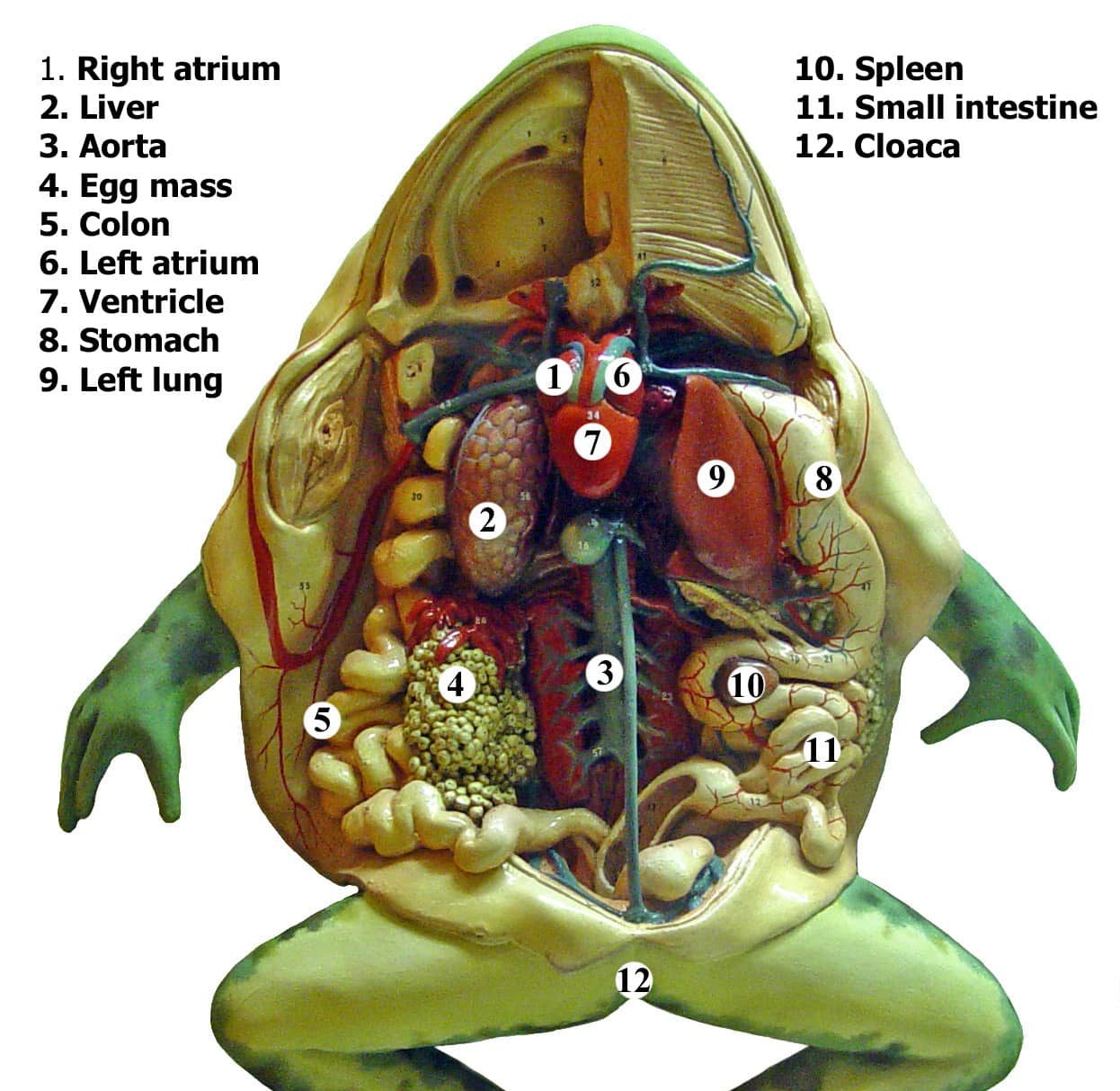
Frog dissection labeled diagram lomishelf
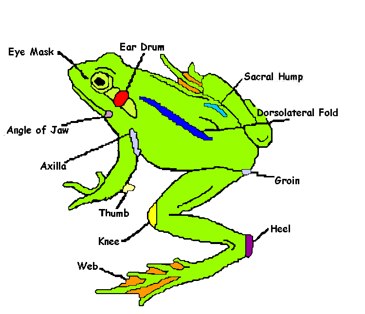
Frog Dissection External Anatomy
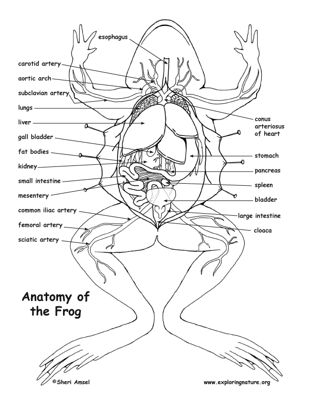
Frog Dissection Diagram and Labeling

The Frog's Anatomy Illustration Poster Graphic poster
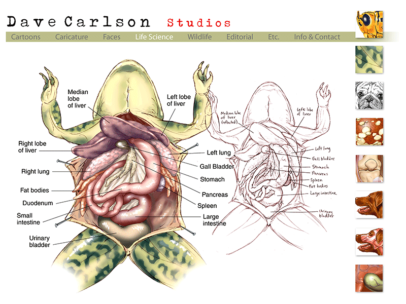
Anatomy Of A Frog

Frog Anatomy, Illustration Stock Image C030/7183 Science Photo

Frog Dissection MRS. MERRITT'S BIOLOGY CLASS

Frog Dissection Art Print by turtletimehewitt XSmall in 2021 Frog

Frog Dissection Carolina Biological Supply
Along With Toads, They Make Up The Largest Group Of Amphibians.
Along With Toads, They Make Up The Largest Group Of Amphibians.
Web Obtain A Preserved Frog And Place It On Your Dissecting Tray, Dorsal Surface Up.
Web Opal Art 5.89K Subscribers Subscribe 2.7K Views 1 Year Ago 10Th Biology Federal Board Fsc, Biology Practical Copy, Experiment 25 Indentification Of Different Parts Of The Respiratory And.
Related Post: